Fungal Diversity Assessment: Aeromycoflora of Indoor And Outdoor Environments at Central University of Kashmir Life Sciences Campus
-
Shaiesta Shah


Department of Plant Sciences, School of Life Sciences, Central University of Kashmir, Nunar, Jammu and Kashmir
Ayman AltafDepartment of Plant Sciences, School of Life Sciences, Central University of Kashmir, Nunar, Jammu and Kashmir
| Received 06 Jan, 2025 |
Accepted 27 May, 2025 |
Published 31 Dec, 2025 |
Background and Objective: Airborne fungal spores significantly impact environmental and human health, acting as allergens and potential sources of mycotoxins. However, there is a paucity of systematic studies quantifying fungal diversity in indoor and outdoor environments within institutional settings. This study evaluates the diversity and abundance of fungal species in indoor and outdoor environments at the Life Sciences Campus, Central University of Kashmir, Nunar, Jammu and Kashmir using standardized sampling and analytical techniques. Materials and Methods: The study, conducted from March to September, 2022, investigated airborne fungal diversity at 15 locations on the Life Sciences Campus, using the gravity plate method with 65 Petri plates. ANOVA was applied to assess significant differences in fungal abundance and diversity between indoor and outdoor sites, with a significance threshold of p<0.05. Results: Predominance of Alternaria alternata, constituting 21.90% of the fungal colonies, followed closely by Aspergillus spp. (12.39%) and Mucor spp. (12.37%). Other notable fungi included Saccharomyces spp., Penicillium spp. and Rhizopus spp. The analysis also revealed that 6.62% of the colonies were sterile hyphae and Blastomyces spp. was the least common. Additionally, dust mites were observed in older cultures, emphasizing the interconnectedness of fungi, dust mites and their environments. Conclusion: This study provides crucial insights into the fungal ecology of the campus, contributing to a better understanding of airborne fungal communities and their potential impacts.
| Copyright © 2025 Shah and Altaf. This is an open-access article distributed under the Creative Commons Attribution License, which permits unrestricted use, distribution, and reproduction in any medium, provided the original work is properly cited. |
INTRODUCTION
Aeromycoflora comprises airborne fungi that significantly influence environmental and human health. Airborne fungal spores, a major component of bioaerosols, include an estimated 80,000 globally distributed species1. These spores are a subject of aero microbiology, which investigates microorganisms’ transport, dispersal and impacts, including fungi, pollen grains and other aerospora. Aerobiology complements this focus by studying the passive movement of biological particles through the atmosphere, emphasizing their sources, deposition and implications for ecosystems and human health2,3. The interplay between indoor and outdoor fungal communities is particularly relevant due to the unique environmental conditions and their potential impacts on air quality.
Airborne fungi are typically dispersed through aerosolization, which may occur naturally via wind or anthropogenic activities such as construction, agriculture, or industrial processes4. Once airborne, fungal spores can travel significant distances and remain viable for extended periods. Several factors, including seasonal variations, time of day, geographic location and environmental settings influence their abundance and diversity1. For instance, fungal spores are more prevalent during warmer, humid seasons such as spring and summer, which promote fungal growth and spore release. Temporal patterns reveal higher spore concentrations during the early morning when cooler temperatures and elevated humidity favor dispersal. Geographic differences also play a role, with coastal and humid regions generally exhibiting higher airborne fungal concentrations compared to arid regions5,6. The health implications of aeromycoflora are profound, as fungal spores can act as potent allergens and cause adverse health effects. A llergic conditions such as bronchial asthma, allergic rhinitis and atopic dermatitis affect 12-20% of the global population, often triggered by fungal spore concentrations exceeding 106 spores/m3 7,8. Furthermore, many fungal species produce secondary metabolites, such as aflatoxins from Aspergillus flavus, which are highly toxic, mutagenic and carcinogenic8. The interaction between fungal biodiversity and environmental factors underscores the need for systematic monitoring, particularly in enclosed environments like educational institutions, where exposure risks may be heightened.
This study aimed to systematically quantify the indoor and outdoor aeromycoflora of the Life Sciences Campus at the Central University of Kashmir, Nunar, Jammu and Kashmir Ganderbal. By addressing these aspects, the study provides valuable insights into fungal ecology and its implications for air quality management in institutional settings.
MATERIALS AND METHODS
Study area: This study was conducted to analyze the diversity and distribution of airborne fungi (aeromycoflora) within the School of Life Sciences Campus, Central University of Kashmir, Nunar, Jammu and Kashmir over seven months (March-September, 2022).
Site selection: Fifteen sites were strategically selected to represent diverse microenvironments within the campus. These included 12 indoor sites, such as classrooms, laboratories, staff rooms, the library, canteen and 3 outdoor locations: The front lawn, mulberry tree beds and the campus backyard. The selection was based on factors like human activity, ventilation levels and proximity to potential fungal sources (e.g., vegetation or damp areas). These sites were chosen to ensure a comprehensive sampling of areas with varying conditions likely to influence fungal diversity.
Sampling procedure: Sampling was carried out over seven months (March to September, 2022) to account for temporal variations in fungal abundance. Samples were collected twice a day, in the morning (9-11 am) and noon (12-2 pm), to capture potential diurnal fluctuations.
The gravity plate method9,10 was used for sampling. Sterile Petri plates containing potato dextrose agar (PDA) supplemented with rifamycin (0.05 g/L) to inhibit bacterial contamination were exposed to ambient air at a height of 1 m for 10 min. After exposure, the plates were sealed with parafilm and transported to the laboratory for incubation at 28±2°C for 7-10 days.
Fungal identification: Fungal colonies growing on the PDA plates were examined for morphological characteristics such as spore shape, hyphal structure, septation and pigmentation. Identification was performed using standard fungal identification manuals11. Dominant species were recorded and colony co65TRGF VuBX nts were expressed as percentages relative to the total number of colonies. The fungi were identified up to the genus level and, in some cases, up to the species level12.
The total occurrence was calculated using the following standard formula13:
 |
Statistical analysis: To ensure the study’s methodological rigor, statistical analysis were employed. The Shannon-Wiener Index was calculated to measure fungal diversity at each site. One-way Analysis of Variance (ANOVA) was used to test for significant differences in fungal abundance and diversity between indoor and outdoor sites. A significance threshold of p<0.05 was used. These analysis provided a quantitative basis for interpreting the results and validating conclusions.
RESULTS
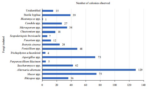
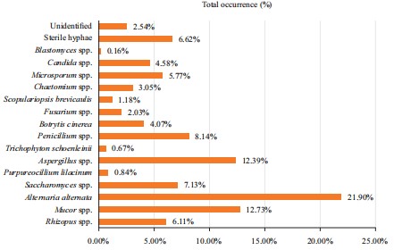
Fungal diversity and dominant species: A total of 65 Petri plates collected from 15 locations yielded a diverse assemblage of 15 fungal genera, highlighting significant variation across indoor and outdoor environments. The most dominant fungal species was Alternaria alternata, which accounted for 21.90% (129 colonies) of the total isolates, followed by Mucor spp. (12.73%, 75 colonies) and Aspergillus spp. (12.39%, 73 colonies) (Fig. 1 and 2).
|
|
|
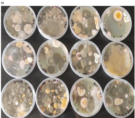
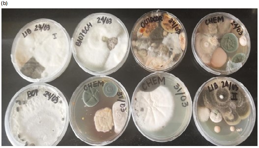
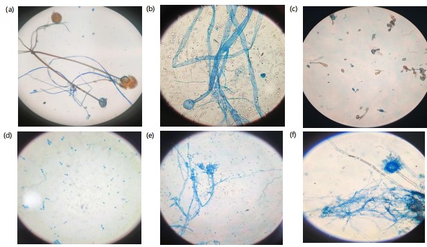
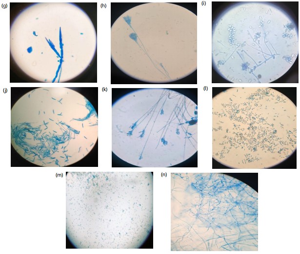
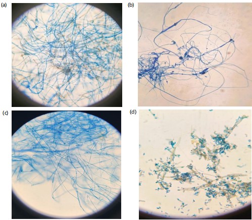
Other frequently observed species included Penicillium spp. (8.14%, 48 colonies), Saccharomyces spp. (7.13%, 42 colonies) and sterile hyphae (6.62%, 39 colonies), suggesting the presence of non-sporulating fungi. Rarely detected species included Scopulariopsis brevicaulis (1.18%, 7 colonies), Purpureocillium lilacinum (0.84%, 5 colonies) and Blastomyces spp. (0.16%, 1 colony), indicating their limited distribution within the sampled environments. The colony characteristics of all the fungi isolated from various locations differ to a greater extent (Fig. 3a-b). The micrographs of a few identified fungi isolated are represented in (Fig. 4a-n) and the unidentified fungi are represented in (Fig. 5a-d).
|
|
Indoor vs outdoor fungal distribution:
| • | Outdoor environments exhibited higher fungal diversity compared to indoor settings, as indicated by the Shannon-Wiener Index values (2.75 outdoors vs 1.89 indoors) | |
| • | Alternaria alternata was the most abundant species in outdoor locations, with mulberry tree beds (21.70%) and the campus backyard (24.03%) showing the highest occurrences | |
| • | In contrast, indoor environments were characterized by the dominance of Aspergillus spp., particularly in the canteen (19.84%) and the botany staffroom (20.75%), likely due to high humidity and organic matter availability |
Temporal variations in fungal abundance:
| • | Morning samples exhibited significantly higher fungal diversity than those collected at noon, likely due to favorable humidity, temperature and reduced human activity during early hours | |
| • | Species such as Alternaria alternata and Mucor spp. were most abundant during morning collections, particularly in outdoor locations where environmental conditions facilitated sporulation and dispersal | |
| • | Rare species, including Scopulariopsis brevicaulis and Blastomyces spp., were predominantly observed in morning samples, suggesting their sensitivity to microclimatic changes throughout the day | |
| • | This study provides valuable insights into the distribution and dynamics of airborne fungi, emphasizing the need for continuous monitoring in institutional settings to assess potential health risks and improve indoor air quality |
DISCUSSION
The study investigated the diversity and distribution of airborne fungi across indoor and outdoor environments within the School of Life Sciences Campus, Central University of Kashmir, Nunar, Jammu and Kashmir. The findings provide valuable insights into fungal prevalence, environmental factors influencing their distribution and implications for health and indoor air quality.
The dominance of Alternaria alternata (21.90%) in this study aligns with findings by Al-Suwaine et al.14, who reported its prevalence in educational and healthcare settings. Alternaria alternata is known for its adaptability to diverse environmental conditions and its role as an aeroallergen, capable of triggering respiratory disorders such as asthma15. Similarly, the frequent occurrence of Aspergillus spp. (12.39%) corroborates the findings of Kim et al.16, which highlighted its abundance in poorly ventilated indoor environments. These genera thrive in organic-rich and damp conditions, as evidenced in this study’s canteen and staff room samples.
Outdoor fungal diversity was significantly higher than indoors, as shown by the Shannon-Wiener Index (2.75 vs 1.89). This aligns with Jones and Harrison17, who reported that outdoor fungal populations are influenced by vegetation, soil activity and climatic conditions. The high concentrations near mulberry tree beds and the campus backyard reflect the role of vegetation in promoting fungal sporulation18. These areas likely acted as reservoirs for spore dispersal into adjacent indoor spaces.
The findings underscore potential health risks associated with prolonged exposure to airborne fungi. Indoor environments, particularly those with limited ventilation, showed a higher prevalence of Aspergillus spp., which is known to cause aspergillosis in immuno-compromised individuals19. Similarly, Cladosporium spp., although less dominant, has been implicated in allergic rhinitis and hypersensitivity pneumonitis. The study highlights the importance of maintaining indoor air quality in educational institutions to safeguard the health of students and staff.
The study employed the gravity plate method, which is widely used for fungal sampling due to its simplicity and cost-effectiveness20. However, it has limitations, including its inability to capture smaller fungal spores and underestimating fungal diversity in low-spore-density environments. Additionally, morphological identification, while effective for common genera, may lead to misclassification, especially for closely related species. Future studies should integrate molecular techniques such as DNA barcoding to enhance accuracy21.
The statistical analysis employed (ANOVA) strengthened the study by confirming significant differences in fungal abundance across sites. The use of diversity indices provided a quantitative framework for comparing fungal populations, adding rigour to the findings. However, the study could benefit from multivariate analysis to explore correlations between fungal abundance and environmental parameters such as humidity and temperature.
Environmental factors play a crucial role in shaping fungal diversity. The high outdoor fungal concentrations can be attributed to vegetation cover, soil microbial activity and higher exposure to environmental fluctuations. Conversely, indoor environments exhibited reduced diversity but dominance of opportunistic fungi like Aspergillus spp., likely due to limited air circulation and human activities. Temporal trends, with higher diversity observed in morning samples, suggest that dew and reduced human interference facilitate fungal growth and sporulation.
The findings have implications beyond educational institutions. Airborne fungal monitoring is critical in healthcare facilities, residential spaces and workplaces to mitigate health risks and improve environmental hygiene. By providing evidence-based recommendations, this study contributes to the growing field of indoor air quality management and fungal ecology.
CONCLUSION
This study provides a comprehensive analysis of airborne fungal diversity in the School of Life Sciences Campus, Central University of Kashmir, Nunar, Jammu and Kashmir. Key findings include the dominance of Alternaria alternata, Aspergillus spp. and Mucor spp., with outdoor locations exhibiting significantly higher diversity than indoor environments. These results emphasize the need for regular monitoring and targeted interventions to maintain indoor air quality and safeguard public health.
Practical recommendations derived from this study include improving ventilation systems, implementing rigorous cleaning protocols and conducting periodic fungal monitoring. Future research should focus on exploring seasonal variations and using advanced molecular methods to enhance our understanding of airborne fungi in educational and public spaces.
SIGNIFICANCE STATEMENT
This study identified the dominance of Alternaria alternata, Aspergillus spp. and Mucor spp. in indoor and outdoor environments, which could be beneficial for understanding fungal distribution patterns and their potential health implications. The findings provide valuable insights into environmental factors influencing fungal diversity and highlight the importance of maintaining air quality in educational institutions. This study will assist researchers in uncovering critical areas of airborne fungal ecology, particularly in understudied educational settings, that have remained unexplored by many. Consequently, a new theory on the interplay between environmental factors and fungal prevalence in indoor and outdoor spaces may be developed.
REFERENCES
- Kerssies, A., 1993. Horizontal and vertical distribution of airborne conidia of Botrytis cinerea in a gerbera crop grown under glass. Neth. J. Plant Pathol., 99: 303-311.
- Burge, H.A. and C.A. Rogers, 2000. Outdoor allergens. Environ. Health Perspect., 108: 653-659.
- Burge, H.A., 2001. Fungi: Toxic killers or unavoidable nuisances? Ann. Allergy Asthma Immunol., 87: 52-56.
- Wang, I.J., Y.L. Guo, H.J. Weng, W.S. Hsieh, Y.L. Chuang, S.J. Lin and P.C. Chen, 2007. Environmental risk factors for early infantile atopic dermatitis. Pediatr. Allergy Immunol., 18: 441-447.
- Khan, A.A.H. and S.M. Karuppayil, 2012. Fungal pollution of indoor environments and its management. Saudi J. Biol. Sci., 19: 405-426.
- Perdelli, F., M.L. Cristina, M. Sartini, A.M. Spagnolo and M. Dallera et al., 2006. Fungal contamination in hospital environments. Infect. Control Hosp. Epidemiol., 27: 44-47.
- Terui, T., Y. Makino, M. Okada, A. Hashimoto and H. Tagami, 2000. Learning from fungus allergy in atopic dermatitis patients [In Japanese]. Jpn. J. Med. Mycol., 41: 157-160.
- Akiyama, K., 2001. Fungal allergy-A clinical perspective [In Japanese]. Jpn. J. Med. Mycol., 42: 109-111.
- Asan, A., S. Ilhan, B. Sen, I.P. Erkara and C. Filik et al., 2004. Airborne fungi and actinomycetes concentrations in the air of Eskisehir City (Turkey). Indoor Built Environ., 13: 63-74.
- Naim Uddin, 2004. Airspora studies over a rice (high yielding variety) field in rabi season in the state of West Bengal, India. Aerobiologia, 20: 127-134.
- Barnett, H.L. and B.B. Hunter, 1972. Illustrated Genera of Imperfect Fungi. 3rd Edn., Burgess Publishing Company, Minneapolis, Minnesota, ISBN: 9780808702665, Pages: 241.
- Cooke, W.B., 1963. A Laboratory Guide to Fungi in Polluted Waters, Sewage, and Sewage Treatment Systems: Their Identification and Culture. Division of Water Supply and Pollution Control, Cincinnati, Ohio, Pages: 132.
- Abdel Hameed, A.A., M.I. Khoder, H.Y. Ibrahim, Y. Saeed, E.M. Osman and S. Ghanem, 2012. Study on some factors affecting survivability of airborne fungi. Sci. Total Environ., 414: 696-700.
- Al-Suwaine, A.S., A.H. Bahkali and S.M. Hasnain, 1999. Seasonal incidence of airborne fungal allergens in Riyadh, Saudi Arabia. Mycopathologia, 145: 15-22.
- Abel-Fernández, E., M.J. Martínez, T. Galán and F. Pineda, 2023. Going over fungal allergy: Alternaria alternata and its allergens. J. Fungi, 9.
- Kim, K.H., E. Kabir and S.A. Jahan, 2018. Airborne bioaerosols and their impact on human health. J. Environ. Sci., 67: 23-35.
- Jones, A.M. and R.M. Harrison, 2004. The effects of meteorological factors on atmospheric bioaerosol concentrations-A review. Sci. Total Environ., 326: 151-180.
- Chawla, H., P. Anand, K. Garg, N. Bhagat and S.G. Varmani et al., 2023. A comprehensive review of microbial contamination in the indoor environment: Sources, sampling, health risks, and mitigation strategies. Front. Public Health, 11.
- Kwon-Chung, K.J. and J.A. Sugui, 2013. Aspergillus fumigatus-What makes the species a ubiquitous human fungal pathogen? PLoS Pathog., 9.
- Savino, E. and G. Caretta, 1992. Airborne fungi in an Italian rice mill. Aerobiologia, 8: 267-275.
- Hibbett, D.S., M. Binder, J.F. Bischoff, M. Blackwell and P.F. Cannon et al., 2007. A higher-level phylogenetic classification of the Fungi. Mycol. Res., 111: 509-547.
How to Cite this paper?
APA-7 Style
Shah,
S., Altaf,
A. (2025). Fungal Diversity Assessment: Aeromycoflora of Indoor And Outdoor Environments at Central University of Kashmir Life Sciences Campus. Asian Journal of Biological Sciences, 18(4), 729-737. https://doi.org/10.3923/ajbs.2025.729.737
ACS Style
Shah,
S.; Altaf,
A. Fungal Diversity Assessment: Aeromycoflora of Indoor And Outdoor Environments at Central University of Kashmir Life Sciences Campus. Asian J. Biol. Sci 2025, 18, 729-737. https://doi.org/10.3923/ajbs.2025.729.737
AMA Style
Shah
S, Altaf
A. Fungal Diversity Assessment: Aeromycoflora of Indoor And Outdoor Environments at Central University of Kashmir Life Sciences Campus. Asian Journal of Biological Sciences. 2025; 18(4): 729-737. https://doi.org/10.3923/ajbs.2025.729.737
Chicago/Turabian Style
Shah, Shaiesta, and Ayman Altaf.
2025. "Fungal Diversity Assessment: Aeromycoflora of Indoor And Outdoor Environments at Central University of Kashmir Life Sciences Campus" Asian Journal of Biological Sciences 18, no. 4: 729-737. https://doi.org/10.3923/ajbs.2025.729.737

This work is licensed under a Creative Commons Attribution 4.0 International License.