
Haematological Biomarkers for Distinguishing Vaso-Occlusive Crisis from Steady State in Patients with Sickle Cell Disease
-
Chinedu Emmanuel Onuoha


Department of Haematology and Transfusion Science, Faculty of Medical Laboratory Science, Federal University, Otuoke, Bayelsa, Nigeria
Vivian Akudo MadukaDepartment of Haematology and Transfusion Science, Faculty of Medical Laboratory Science, Federal University, Otuoke, Bayelsa, Nigeria
Benjamin Onyema EledoDepartment of Haematology and Transfusion Science, Faculty of Medical Laboratory Science, Federal University, Otuoke, Bayelsa, Nigeria
| Received 31 Aug, 2026 |
Accepted 10 Oct, 2026 |
Published 31 Dec, 2026 |
Background and Objective: Vaso-occlusive crisis (VOC) is a major complication of sickle cell disease (SCD), characterized by increased haemolysis, inflammation, and thrombo-inflammatory activity. This study evaluated the diagnostic utility of routine haematological biomarkers for distinguishing patients with SCD during VOC from those in steady state. Materials and Methods: A comparative study was conducted among 180 participants aged 5-40 years in Bayelsa State, Nigeria, comprising 67 patients with SCD in crisis and 53 patients in steady state. Three millilitres of venous blood were collected into EDTA containers and analysed using a five-part haematology analyser. Haematological parameters, including Packed Cell Volume (PCV), White Blood Cell Count (WBC), differential leukocyte counts, Mean Cell Volume (MCV), Mean Cell Haemoglobin (MCH), Mean Cell Haemoglobin Concentration (MCHC), Platelet Count, Mean Platelet Volume (MPV), Absolute Neutrophil Count (ANC), and Neutrophil-to-Lymphocyte Ratio (NLR), were assessed. Data were analysed using SPSS version 27.0, with p<0.05 considered statistically significant. Results: Compared with steady-state patients, those in VOC had significantly lower PCV (21.56±4.54% vs 24.95±2.52%; p=0.007), higher WBC count (12.88±5.65 vs 7.74±2.07×10⁹/L; p = 0.001), neutrophil percentage (62.39±12.87% vs 54.45±5.83%; p=0.017), ANC (7.01±4.52 vs 2.35±0.74×10 /L; p<0.001), and NLR (2.72±1.52 vs 1.52±0.43; p=0.002). Other erythrocyte and platelet indices showed no statistically significant differences. Conclusion: Routine CBC-derived biomarkers, particularly PCV, WBC count, neutrophil percentage, ANC, and NLR, demonstrate potential utility for distinguishing VOC from steady state in SCD. Their accessibility and affordability make them valuable for disease monitoring and clinical decision-making in resource-limited settings.
| Copyright © 2026 Onuoha et al. This is an open-access article distributed under the Creative Commons Attribution License, which permits unrestricted use, distribution, and reproduction in any medium, provided the original work is properly cited. |
INTRODUCTION
Sickle cell disease (SCD) is one of the most common genetic haemoglobin abnormalities in the world and continues to be a major public health concern, particularly in Sub-Saharan Africa. A single nucleotide alteration in the β-globin gene causes aberrant hemoglobin S (HbS) synthesis. A HbS polymerizes in response to hypoxia, dehydration, acidity, or oxidative stress, leading red blood cells to become hard, sickle-shaped, and less deformable. These aberrant erythrocytes clog the microvasculature, causing persistent hemolysis, repeated vaso-occlusion, tissue ischemia, progressive organ damage, and chronic anaemia1,2.
Every year, between 300,000 and 400,000 infants worldwide are born with sickle cell disease; about 75% of these births take place in Sub-Saharan Africa. With almost 150,000 births with sickle cell disease (SCD) each year, Nigeria has the largest global burden of the disease. Sickle cell disease still significantly contributes to childhood mortality, frequent hospitalization, lower quality of life, and early death despite advancements in birth screening, vaccination, hydroxyurea medication, and comprehensive supportive care3,4.
Sickle cell illness has a clinical course that alternates between steady state and vaso-occlusive crisis (VOC). The steady state is a clinically stable time in which patients do not experience acute painful episodes, infections, blood transfusions, or hospitalization for several weeks. Vaso-occlusive crisis, on the other hand, is an immediate complication marked by extreme pain due to vascular obstruction induced by sickled erythrocytes, activated leukocytes, platelets, and endothelial cells. These occurrences are followed with increased inflammation, haemolysis, oxidative stress, and endothelial dysfunction, making VOC the primary cause of hospitalization among sickle cell disease patients5,6.
The role of inflammation in the pathophysiology of vaso-occlusive crisis is becoming more widely acknowledged. In order to promote vascular occlusion and intensify inflammatory reactions, activated neutrophils cling to the vascular endothelium and interact with sickled erythrocytes and platelets. As a result, a number of standard hematological parameters, such as platelet count, Neutrophil-To-Lymphocyte Ratio (NLR), total White Blood Cell (WBC) count, neutrophil count, Absolute Neutrophil Count (ANC), erythrocyte indices, and Neutrophil-To-Lymphocyte Ratio (NLR), have become potential indicators of disease activity and severity6,7.
In the treatment of sickle cell disease, routine Complete Blood Count (CBC) analysis is still one of the most easily accessible, reasonably priced, and educational laboratory tests. Important data on erythropoiesis, hemolysis, inflammatory state, immunological activation, and thrombopoiesis can be found in parameters derived from automated haematology analyzers. These indicators help physicians track the course of an illness, assess how well a treatment is working, spot problems, and make therapeutic decisions. Routine haematological indices are still useful tools for clinical evaluation because advanced molecular and inflammatory biomarkers are frequently unavailable in many impoverished nations8.
Despite numerous studies on haematological abnormalities in sickle cell disease, most have evaluated individual laboratory parameters or compared patients with healthy controls. Evidence on the combined diagnostic utility of routine haematological biomarkers for distinguishing vaso-occlusive crisis from steady state, particularly among Nigerian patients, remains limited. This knowledge gap hinders the effective use of readily available laboratory indices for disease monitoring and clinical decision-making. Identifying reliable, inexpensive, and accessible biomarkers would facilitate early diagnosis, prompt intervention, improved risk stratification, and better clinical outcomes, especially in resource-limited settings.
Therefore, the aim of this study is to evaluate the diagnostic utility of routine haematological biomarkers in distinguishing patients with sickle cell disease in vaso-occlusive crisis from those in steady state.
MATERIALS AND METHODS
Study area: For this study, communities in Bayelsa State with coordinates of 4.8678°N, 5.8987°E were used. Situated between Delta State and River State in the center Niger Delta Region, Bayelsa State is a state in Southern Nigeria with a total surface area of approximately 10,773 km2 (4,159 sq mi). Its capital, Yenagoa, is situated at 4°55'29"N, 6°15'51"E. Although Ijaw is the most widely spoken language, English is the official language. The state was created in 1996 from a section of Rivers State and has an estimated population of 1,704,515 according to the 2006 census. Eight local government areas make up Bayelsa State, which has Yenagoa as its capital. The study was conducted from June 2025 to February 2026.
Study population: The study included 120 male and female volunteers between the ages of 5 and 40. During the study period, 53 stable sickle cell subjects and 67 sickle cell crisis patients were admitted to or attended clinic days at Federal Medical Center Yenagoa, Kaiama General Hospital, Diete Koki Memorial Hospital, and Niger Delta University Teaching Hospital Okolobiri.
Selection criteria: This was done using questionnaire and consent form.
Inclusion criteria: Participants included in the study were confirmed sickle cell patients, both male and female, aged 5 to 40 years, as verified through laboratory screening.
Exclusion criteria: The study excludes:
| • | Sickle cell patients with co-morbidities, including hypertension, diabetes mellitus, chronic kidney disease, and known coagulation disorders | |
| • | Individuals who refused to provide informed consent |
Sample size: According to reports, the South-South region of Nigeria has a 3.7% prevalence of sickle cell disease9. The formula suggested by Bolarinwa10:
was used to determine the sample size.
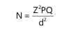 |
Where:
| N | = | Required sample size | |
| Z2 | = | Critical value at 95% confidence level = 1.96 | |
| P | = | Estimated prevalence = 0.037 | |
| Q | = | 1-P = 0.963 | |
| d2 | = | Absolute sampling error tolerated = 0.05 |
The calculations were done as follows:
| Z2 | = | 1.962 = 3.8416 | |
| P | = | 3.7% = 0.037 | |
| Q | = | 1-0.037 = 0.963 | |
| d2 | = | 5% = 0.05 = 0.0025 | |
| N | = | 3.8416×0.037×0.963/0.0025 = 54.7 |
The minimum sample size for this study was 55.
Using 10% attrition rate, the sample size increased to 60 participants each.
Collection of sample: For haematological analysis, 3 mL of venous blood was drawn aseptically using a 5 mL syringe and transferred into an EDTA (Ethylenediaminetetraacetic Acid) tube. To prevent hemolysis and platelet disintegration, analysis was performed using whole blood within two hours of sample collection.
| Table 1: | Haematological parameters between sickle cell subjects in crisis and stable state | |||
| Parameter (Unit) | Crisis (N = 67) | Stable state (N = 53) | t-value | p-value |
| Packed cell volume (%) | 1.56±4.54 | 24.95±2.52 | 2.89 | 0.007 |
| White blood cell count (×109/L) | 12.88±5.65 | 7.74±2.07 | 3.8 | 0.001 |
| Neutrophils (%) | 62.39±12.87 | 54.45±5.83 | 2.49 | 0.017 |
| Lymphocytes (%) | 31.89±12.34 | 39.10±10.47 | 1.95 | 0.059 |
| Monocytes (%) | 3.17±2.12 | 2.70±1.22 | 0.84 | 0.405 |
| Eosinophils (%) | 2.56±0.86 | 2.15±0.99 | 1.35 | 0.187 |
| Basophils (%) | 0.41±0.12 | 0.39±0.10 | 0.56 | 0.58 |
| Mean cell volume (fL) | 74.28±8.34 | 6.19±9.19 | 0.67 | 0.508 |
| Mean cell haemoglobin (pg) | 21.76±4.04 | 21.41±2.14 | 0.34 | 0.737 |
| Mean cell haemoglobin conc (g/dL) | 42.79±63.52 | 25.97±2.42 | 1.19 | 0.243 |
| Platelet Count (×109/L) | 411.72±137.04 | 340.40±103.55 | 1.82 | 0.077 |
| Absolute neutrophil count (×109/L) | 7.01±4.52 | 2.35±0.74 | 4.55 | 0.001 |
| Mean platelet volume (fL) | 10.68±0.46 | 11.24±1.18 | 1.87 | 0.07 |
| Neutrophil-Lymphocyte ratio (Unitless) | 2.72±1.52 | 1.52±0.43 | 3.41 | 0.002 |
| Values are presented as Mean±Standard deviation (SD). Statistical comparisons were performed among the various study groups, and a p-value<0.05 was considered statistically significant | ||||
Ethical approval: The Bayelsa State Ministry of Health, which oversees all Bayelsa State health facilities, provided ethical clearance, as did the Federal Medical Center Yenagoa research committee. Respect for participants’ rights was observed, including the right to refuse participation with an explanation provided on the participant information form.
Statistical analysis: The Statistical Package for the Social Sciences (SPSS) version 27.0 was used to enter, clean, and analyze the data. A p-value of less than 0.05 was deemed statistically significant, and continuous data were reported as mean±standard deviation (SD). The independent samples t-test was used to examine the differences between crisis and stable.
Method of laboratory analysis: Haematological Parameterswere done by 5-part Haematology Analyzer (wincom HA8520)while the electrophoresis were performed using the Model Techmel and Techmel 300 (USA) systems.
RESULTS
Table 1 presents a comparison of selected haematological parameters between sickle cell subjects in crisis and those in the stable state, in line with the study objective of evaluating haematological alterations associated with disease activity.
The packed cell volume (PCV) was significantly lower among sickle cell subjects in crisis (21.56±4.54%) compared with those in the stable state (24.95±2.52%), with a statistically significant difference (p = 0.007). The white blood cell count was significantly elevated in subjects in crisis (12.88±5.65×10⁹/L) compared with those in the stable state (7.74±2.07×10⁹/L), with a highly significant difference (p = 0.001). Neutrophil percentage was significantly higher in crisis subjects (62.39±12.87%) than in those in the stable state (54.45±5.83%), with a statistically significant difference (p = 0.017). Lymphocyte percentage was lower among crisis subjects (31.89±12.34%) compared with stable-state subjects (39.10±10.47%); however, this difference did not reach statistical significance (p = 0.059).
Red cell indices, including mean cell volume, mean cell haemoglobin, and mean cell haemoglobin concentration, did not differ significantly between crisis and stable-state subjects (p>0.05).
Platelet count was higher among crisis subjects (411.72±137.04×10⁹/L) compared with stable-state subjects (340.40±103.55×10⁹/L), although this difference did not reach statistical significance (p = 0.077). The absolute neutrophil count was markedly higher in crisis subjects (7.01±4.52×10⁹/L) compared with stable-state subjects (2.35±0.74×10⁹/L), with a highly significant difference (p<0.001). Mean platelet volume was slightly lower in crisis subjects (10.68±0.46 fL) compared with stable-state subjects (11.24±1.18 fL), but the difference was not statistically significant (p = 0.070).
The neutrophil-lymphocyte ratio was significantly higher in crisis subjects (2.72±1.52) than in stable-state subjects (1.52±0.43), with a statistically significant difference (p = 0.002).
DISCUSSION
Erythrocyte abnormalities continue to be essential clinical events that differentiate vaso-occlusive crises from the steady state, as shown by the combined interpretation of Table 1. Rapid intravascular and extravascular hemolysis brought on by recurrent erythrocyte sickling, membrane instability, oxidative damage, and shorter erythrocyte lifespan is reflected in the notable decrease in packed cell volume during a crisis. In the end, these mechanisms lower oxygen delivery and cause tissue hypoxia, which prolongs vaso-occlusion1,5.
Interestingly, although MCV and MCH were not statistically different between groups, their graphical trends show that erythropoiesis is impaired throughout the disease course. This implies that erythrocyte indices may reflect chronic bone marrow adaptation, whereas PCV responds faster to acute haemolytic events. Similarly, the considerably greater MCHC found during VOC supports increased erythrocyte dehydration, which is a well-established mechanism that promotes intracellular HbS polymerization and cellular stiffness6.
The integrated use of several erythrocyte parameters rather than relying just on PCV or hemoglobin content is a noteworthy innovation of this study. The picture depicts the range of erythrocyte damage throughout the course of the illness, emphasizing how routine CBC-derived parameters as a whole offer more diagnostic information than individual variables. In situations where sophisticated hemolysis biomarkers such plasma free hemoglobin, lactate dehydrogenase, and erythrocyte deformability assays are unavailable, this integrated interpretation provides clinicians with an easy and cost-effective way to track hemolytic severity11,12.
The leucocyte profile revealed in this study lends support to the emerging theory that sickle cell disease is basically a chronic inflammatory condition characterized by bouts of excessive innate immune activation. During VOC, the WBC count, neutrophil percentage, ANC, and NLR levels were significantly higher, indicating that neutrophil-mediated inflammatory pathways had been activated. Activated neutrophils interact with sickled erythrocytes, activated platelets, and endothelial cells, causing endothelial damage, increased expression of adhesion molecules, oxidative stress, and microvascular blockage6,13.
The large increase in ANC is especially significant since absolute neutrophil count directly indicates the extent of inflammatory activation and may be less affected by changes in total leukocyte count than neutrophil percentage alone. Similarly, the considerable rise of NLR indicates both neutrophilia and relative lymphocyte suppression, providing a comprehensive index of inflammatory load. Recent research reveals that NLR predicts hospitalization, acute chest syndrome, recurrent vaso-occlusive crises, and death in SCD patients, making it an appealing prognostic biomarker because it is easily accessible with routine CBC study14,15.
A particularly innovative aspect of this study is the demonstration that routine leukocyte-derived biomarkers can distinguish crisis from steady state without requiring expensive inflammatory assays such as interleukin-6, tumour necrosis factor-alpha, C-reactive protein, or soluble adhesion molecules. This finding has considerable clinical implications for low-resource healthcare systems, where CBC remains the most accessible laboratory investigation.
Furthermore, the graphical representation provides a visual inflammatory signature of VOC, clearly illustrating progressive immune activation and emphasizing that routine haematological parameters possess substantial diagnostic value when interpreted collectively rather than individually.
Platelet abnormalities observed in this study reinforce the concept that thrombosis and inflammation are inseparable processes in sickle cell disease. Although platelet count was higher during VOC, MPV showed a slight reduction, suggesting increased consumption of larger, metabolically active platelets at sites of vascular occlusion. Activated platelets release pro-inflammatory cytokines, thromboxane A2, platelet factor 4, and express P-selectin, thereby facilitating interactions with neutrophils and endothelial cells that amplify vaso-occlusion16,17.
The coexistence of thrombocytosis with increased neutrophil activity further supports the emerging paradigm that platelet-leukocyte aggregates are central mediators of vaso-occlusive pathology. These aggregates promote endothelial activation, NET formation, and thrombin generation, establishing a self-amplifying cycle of inflammation and coagulation. Consequently, platelet count should no longer be regarded merely as an indicator of thrombopoiesis but also as a biomarker reflecting thrombo-inflammatory activity.
A major innovation of this study is the integration of platelet indices with inflammatory leukocyte biomarkers into a composite CBC-based diagnostic panel. Rather than assessing platelet count in isolation, the study demonstrates that combining platelet count with ANC and NLR substantially improves differentiation between VOC and steady state. This integrated strategy has important translational relevance because it utilizes routinely available laboratory parameters without increasing diagnostic costs.
Collectively, the erythrocyte and leukocyte findings by demonstrating that SCD pathophysiology results from coordinated interactions among haemolysis, inflammation, and thrombosis. This multidimensional interpretation advances current understanding of SCD as a chronic thrombo-inflammatory disease rather than solely a disorder of abnormal haemoglobin.
Routine Complete Blood Count (CBC) evaluation should include erythrocyte, leukocyte, and platelet-derived indices, particularly Packed Cell Volume (PCV), Absolute Neutrophil Count (ANC), Neutrophil-To-Lymphocyte Ratio (NLR), platelet count, and Mean Platelet Volume (MPV), to support comprehensive monitoring of patients with sickle cell disease. Clinicians should consider ANC, NLR, and platelet indices as inexpensive and readily available inflammatory biomarkers for assessing disease activity and identifying patients at increased risk of vaso-occlusive crises and related complications. Healthcare institutions should strengthen access to automated haematology analysers and promote standardized interpretation of CBC-derived biomarkers, particularly in low-resource settings. Future multicentre prospective studies should further evaluate the prognostic value of these haematological biomarkers in predicting hospitalization, recurrent vaso-occlusive crises, acute chest syndrome, organ dysfunction, and mortality. Further research should also integrate routine haematological indices with inflammatory cytokines, endothelial activation markers, coagulation biomarkers, and genetic modifiers to develop robust predictive models for disease severity and individualized management.
This study demonstrates distinct and progressive alterations in erythrocyte, leukocyte, and platelet biomarkers between steady-state sickle cell disease and vaso-occlusive crisis, reflecting the underlying changes associated with disease activity. The findings indicate that routine CBC-derived indices, particularly ANC, NLR, platelet count, and erythrocyte indices, may serve as practical, affordable, and readily available biomarkers for evaluating disease activity. By integrating erythrocyte, leukocyte, and platelet parameters rather than focusing on a single haematological marker, the study provides a broader assessment of the interactions among haemolysis, inflammation, and thrombosis during disease progression. The graphical presentation further complements the tabulated findings by illustrating changes in haematological parameters between steady state and vaso-occlusive crisis. Overall, the findings support the concept of sickle cell disease as a chronic thrombo-inflammatory disorder involving interactions among red blood cells, leukocytes, platelets, and the vascular endothelium. The locally generated evidence also supports the potential incorporation of routine CBC-derived biomarkers into clinical monitoring and provides a basis for future studies investigating predictive and individualized approaches to sickle cell disease management.
CONCLUSION
This study demonstrated significant alterations in erythrocyte, leukocyte, and platelet biomarkers among patients with sickle cell disease, with the greatest abnormalities occurring during vaso-occlusive crisis. Reduced erythrocyte indices reflected worsening haemolytic anaemia, while elevated total white blood cell count, absolute neutrophil count, neutrophil-to-lymphocyte ratio, and platelet count indicated heightened inflammatory and thrombo-inflammatory activity. The findings confirm that sickle cell disease is a multisystem disorder characterized by the interplay of chronic haemolysis, inflammation, and platelet activation. Importantly, routine complete blood count-derived biomarkers provide simple, affordable, and reliable tools for assessing disease activity, distinguishing vaso-occlusive crisis from the steady state, and supporting clinical management, particularly in resource-limited settings.
SIGNIFICANCE STATEMENT
This study is significant because it demonstrates that routine Complete Blood Count (CBC)-derived biomarkersincluding Packed Cell Volume (PCV), White Blood Cell Count (WBC), Absolute Neutrophil Count (ANC), Neutrophil-to-Lymphocyte Ratio (NLR), and platelet indices can reliably distinguish vaso-occlusive crisis from steady state in patients with sickle cell disease. Unlike advanced molecular assays, these parameters are affordable, accessible, and widely available, making them particularly valuable in resource-limited healthcare settings. By integrating erythrocyte, leukocyte, and platelet indices into a unified diagnostic framework, the study provides a novel, practical approach for monitoring disease activity, guiding timely clinical decisions, and improving patient outcomes. This work contributes to the growing understanding of sickle cell disease as a chronic thrombo-inflammatory disorder and establishes routine haematological biomarkers as effective tools for risk stratification and management.
REFERENCES
- Kato, G.J., F.B. Piel, C.D. Reid, M.H. Gaston and K. Ohene-Frempong et al., 2018. Sickle cell disease. Nat. Rev. Dis. Primers, 4.
- Piel, F.B., M.H. Steinberg and D.C. Rees, 2017. Sickle cell disease, N. Engl. J. Med., 376: 1561-1573.
- Rajput, H.S., M. Kumari, C. Talele, C. Sajan, V. Saggu and R. Hadia, 2024. Comprehensive overview of sickle cell disease: Global impact, management strategies, and future directions. J. Adv. Zool., 45: 561-566.
- Yassin, M., C. Minniti, N. Shah, S. Alkindi and F. Ata et al., 2025. Evidence and gaps in clinical outcomes of novel pharmacologic therapies for sickle cell disease: A systematic literature review highlighting insights from clinical trials and real-world studies. Blood Rev., 73.
- Rees, D.C., T.N. Williams and M.T. Gladwin, 2010. Sickle-cell disease. Lancet, 376: 2018-2031.
- Conran, N. and J.D. Belcher, 2018. Inflammation in sickle cell disease. Clin. Hemorheol. Microcirc., 68: 263-299.
- Pavitra, E., R.K. Acharya, V.K. Gupta, H.K. Verma and H. Kang et al., 2024. Impacts of oxidative stress and anti-oxidants on the development, pathogenesis, and therapy of sickle cell disease: A comprehensive review. Biomed. Pharmacother., 176.
- Briggs, C. and B.J. Bain, 2017. Basic Haematological Techniques. In: Dacie and Lewis Practical Haematology, Bain, B.J., I. Bates and M.A. Laffan, Elsevier, Amsterdam, Netherlands, ISBN: 978-0-7020-6696-2, pp: 18-49.
- West, B.A. and J.E. Aitafo, 2023. Prevalence, pattern of disease and outcome of children with sickle cell disease admitted in a private health facility in Southern Nigeria. Asian J. Pediatr. Res., 12: 17-27.
- Bolarinwa, O.A., 2020. Sample size estimation for health and social science researchers: The principles and considerations for different study designs. Niger. Postgrad. Med. J., 27: 67-75.
- Sesti-Costa, R., F.F. Costa and N. Conran, 2023. Role of macrophages in sickle cell disease erythrophagocytosis and erythropoiesis. Int. J. Mol. Sci., 24.
- Bhatt, S., D.A. Argueta, K. Gupta and S. Kundu, 2024. Red blood cells as therapeutic target to treat sickle cell disease. Antioxid. Redox Signaling, 40: 1025-1049.
- Sundd, P., M.T. Gladwin and E.M. Novelli, 2019. Pathophysiology of sickle cell disease. Annu. Rev. Pathol. Mech. Dis., 14: 263-292.
- Buonacera, A., B. Stancanelli, M. Colaci and L. Malatino, 2022. Neutrophil to lymphocyte ratio: An emerging marker of the relationships between the immune system and diseases. Int. J. Mol. Sci., 23.
- Obeagu, E.I. and G.U. Obeagu, 2024. Clinical implications of neutrophil-to-lymphocyte ratio in sickle cell disease. Haematol. Int. J., 8.
- Morrell, C.N., A.A. Aggrey, L.M. Chapman and K.L. Modjeski, 2014. Emerging roles for platelets as immune and inflammatory cells. Blood, 123: 2759-2767.
- Mereweather, L.J., A. Constantinescu-Bercu, J.T.B. Crawley and I.I. Salles-Crawley, 2023. Platelet-neutrophil crosstalk in thrombosis. Int. J. Mol. Sci., 24.
How to Cite this paper?
APA-7 Style
Onuoha,
C.E., Maduka,
V.A., Eledo,
B.O. (2026). Haematological Biomarkers for Distinguishing Vaso-Occlusive Crisis from Steady State in Patients with Sickle Cell Disease. Asian Journal of Biological Sciences, 19(4), 117-124. https://doi.org/10.3923/ajbs.2026.117.124
ACS Style
Onuoha,
C.E.; Maduka,
V.A.; Eledo,
B.O. Haematological Biomarkers for Distinguishing Vaso-Occlusive Crisis from Steady State in Patients with Sickle Cell Disease. Asian J. Biol. Sci 2026, 19, 117-124. https://doi.org/10.3923/ajbs.2026.117.124
AMA Style
Onuoha
CE, Maduka
VA, Eledo
BO. Haematological Biomarkers for Distinguishing Vaso-Occlusive Crisis from Steady State in Patients with Sickle Cell Disease. Asian Journal of Biological Sciences. 2026; 19(4): 117-124. https://doi.org/10.3923/ajbs.2026.117.124
Chicago/Turabian Style
Onuoha, Chinedu, Emmanuel, Vivian Akudo Maduka, and Benjamin Onyema Eledo.
2026. "Haematological Biomarkers for Distinguishing Vaso-Occlusive Crisis from Steady State in Patients with Sickle Cell Disease" Asian Journal of Biological Sciences 19, no. 4: 117-124. https://doi.org/10.3923/ajbs.2026.117.124

This work is licensed under a Creative Commons Attribution 4.0 International License.
