Ameliorative Effect of Tender Coconut Water on Benzene Induced Lymphoid Malignancy in Wistar Rat
-
Onuoha Chinedu Emmanuel


Department of Haematology and Transfusion Science, Faculty of Medical Laboratory Science, Federal University Otuoke, Otuoke, Bayelsa, Nigeria
Ezekiel Fayiah HallieSchool of Pharmacy, University of Liberia, Monrovia, Liberia
| Received 24 Aug, 2023 |
Accepted 12 Oct, 2023 |
Published 31 Dec, 2023 |
Background and Objective: Coconut water is used for the treatment of various forms of cancer. The dose of administration and mechanism of action are not known and much attention has not been given to benzene-induced lymphoid malignancy. This work aimed to evaluate the ameliorative effect of tender coconut water in benzene-induced lymphoid malignancy in Wistar rats to ascertain the potential therapeutic effect of coconut water in this disease condition. Materials and Methods: Lymphoid malignancy was induced intraperitoneal in Wistar rats by administering 0.14 mL kg-1 b.wt., of benzene solution 48 hrs for 4 weeks. General tissue structures, the p53 gene and Bcl-2 were used to confirm the malignant condition. As 10, 20 and 30 mL kg-1 b.wt., of fresh coconut water were given daily via orogastric intubation for 4 weeks for treatment. The data obtained were analyzed by SPSS software. Results: This induction of lymphoid malignancy was successful in Wistar rats. The histology reports in inguinal lymph node, spleen and bone marrow in a positive control group with overexpression of p53 and Bcl-2 cancer biomarkers. However, treatment with 10, 20 and 30 mL kg-1 b.wt., of Malayan green dwarf hybridized immature (HI) coconut water showed restoration of normal histology of the lymph node, spleen and bone marrow with negative nuclear immunohistochemistry reaction of p53 and negative cytoplasmic staining pattern Bcl-2. Conclusion: This study has shown that benzene-induced lymphoids were ameliorated with Malayan green dwarf hybridized immature (HI) coconut water.
| Copyright © 2023 Emmanuel and Hallie. This is an open-access article distributed under the Creative Commons Attribution License, which permits unrestricted use, distribution, and reproduction in any medium, provided the original work is properly cited. |
INTRODUCTION
Cancer is a disease that may affect the entire system of the body that originates from local manifestations and eventually progresses in various processes resulting in neoangiogenesis, rapid proliferation, resisting cell death, local invasion and remote metastasis1.
Lymphoid malignancies are the totality of various malignant complications from bone marrow cells and the lymphatic system origin2. Genetic modification in the bone marrow cell and lymphoid tissue at the external region is the reason for these clonal diseases3.
Coconut water (coconut liquid endosperm) contains many potential therapeutic properties has high nutritional values and is commonly consumed globally because it is refreshing4. Phytochemicals and Phytohormones are available in coconut water have expressed some remarkable anti-aging, anticancer and anticoagulation effects that contributed to the various health benefits by researchers5,6. It was also discovered to possess a potential for boosting the human body’s antioxidant system, regulating hypertension and offering protection against myocardial infarction4.
The International Agency for Research on Cancer (IARC) has classified benzene as a human haematological carcinogen7. There are strong associations between benzene exposure and Lymphoid malignancies supported by epidemiological studies6,8-10.
The aim of this study was to evaluate the ameliorative effect of coconut water in benzene-induced lymphoid malignancy in Wistar rats.
MATERIALS AND METHODS
Study area: The study was carried out in Pharmacology and Medical Laboratory Science Department, Amassoma, Niger Delta University, Bayelsa State, Nigeria from October, 2021 to April, 2022.
Study population: Thirty Wistar rats were bred in Animal House of the Department of Pharmacology, Niger Delta University, Amassoma, Bayelsa State were used in this study.
Ethical approval: College Health Research Ethics Committee, College Of Health Sciences, Niger Delta University, Amassosoma, Bayelsa State, provided ethical clearance and approval.
Benzene solution: Benzene crystallizable 99% extra pure, a product of Loba Chemie PVT LTD, Mumbai, India with product code 0003800500 was used in this research.
Acute toxicity study: The acute toxicity of benzene was estimated in six Wistar rats, divided into 3 groups, two each using the standard method of Lorke11. Each group was given different doses of 0.115, 0.172 and 0.229 mL kg–1 b.wt., respectively. They were kept in the same laboratory conditions and observed for signs of toxicity such as paw-licking, stretching, respiratory stress and mortality for the first 4 hrs and the number of deaths per group was recorded after 24 hrs.
The working doses in this experiment were calculated using the formula:
where, D0 is the highest dose that resulted in no mortality. The D100 is lowest dose that resulted in death.
There were a total of 3 deaths:
Therefore, the Median Lethal Dose (LD50) of the acute toxicity test of Benzene in Wistar rats was found to be 0.14 mL kg–1 b.wt., while coconut water did not show any sign of toxicity at 10, 20 and 30 mL kg–1 11.
Research protocol: Thirty Wistar rats (170-200 g) bred in the Animal House of the Department of Pharmacology, Niger Delta State, Welberfoce Island, Amamasoma, Bayelsa State were used for this study. The animals were allowed to acclimatize in the animal’s house for about 7 days prior to the experiment. They were kept in properly ventilated cages that were cleaned and food replaced daily at a room temperature of about 37°C. The animals were fed with commercially formulated growers marsh and water from the tap. They were randomly selected and divided into 5 groups with each group consisting of 6 rats. Group 1 serves as a negative control and group 2 is a positive control. Group 1 was given water and a normal diet. The remaining groups were intraperitoneally administered 0.14 mL kg–1 b.wt., of benzene 48 hrs for 4 weeks, then fresh coconut water was given daily via orogastric intubation for 4 weeks.
| Group 1: Normal diet+water (Negative control) | |
| Group 2: Induction with Benzene+normal diet (Positive Control) | |
| Group 3: Induction with Benzene+normal diet+coconut water (Dose 1:10 mL kg–1 b.wt.) | |
| Group 4: Induction with Benzene+normal diet+coconut water (Dose 2:20 mL kg–1 b.wt.) | |
| Group 5: Induction with Benzene+normal diet+coconut water (Dose 3:30 mL kg–1 b.wt.) |
Removal of organs: After 4 weeks of coconut water treatment, the rats were anesthetized by placing them in a glass chamber containing cotton wool soaked in chloroform and they were then humanely sacrificed one by one. The lymph node, bone marrow and spleen were harvested and immediately fixed in 10% formal saline solution for histology, p53 and Bcl-2 studies. General tissue structure by Hematoxylin and Eosin staining techniques, p53 and Bcl-2 (Cancer Biomarkers) by Immunohistochemistry.
Fresh hybridized coconuts were harvested in the coconut farm in Opume, Ogbia Local Government of Bayelsa State and were identified as Malayan green dwarf coconut by a Plant scientist with code number UILH/001/508/2020. Qualitative and quantitative Phytochemicals analysis of the coconut water was done using standard method.
Statistical analysis: The data obtained were analyzed by SPSS software version 22.
RESULTS
Qualitative analysis of fresh Malayan green dwarf hybridized immature coconut water with the following bioactive phytochemical substances: Alkaloids, Tannins, Saponins, Terpenoids, Coumarin, Steroids, Flavonoids, Phenolic and Glycosides as shown in Table 1.
| Table 1: | Qualitative analysis of fresh Malayan green dwarf hybridized immature coconut water | |||
| Samples | MGDHICW |
| Saponin | + |
| Tannin | + |
| Phenolics | + |
| Flavonoids | + |
| Terpenoids | + |
| Triterpene- | |
| Coumarin | + |
| Glycosides | + |
| Steroids | + |
| Aikaloid | + |
| Phlobatanin | - |
| Anthocyanin | - |
| Aminoacids | - |
| +: Detected,-: Not detected and MGDHICW: Malayan green dwarf hybridized immature coconut water | |
|
| Table 2: | Quantitative phytochemicals analysis of fresh Malayan green dwarf hybridized immature coconut water | |||
| Phytochemicals (μg/100 g) | Values |
| Alkaloids | 18.60 |
| Tannins | 3.10 |
| Saponins | 0.21 |
| Terpenoids | 9.70 |
| Coumarins | 27.40 |
| Steroids | 149.80 |
| Flavanoids | 48.00 |
| Phenolics | 406.00 |
| Glycosides | 7.00 |
| N | 7.00 |
| Mean±SD | 74.42 ±132.75 |
Quantitative Phytochemical analysis of fresh Malayan green dwarf hybridized immature coconut water with a mean value of bioactive phytochemical substances: 74.42±132.75 as shown in Table 2.
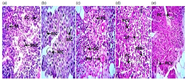
Figure 1(a-e) shows (a) Negative control showing normal histology of the lymph node with cortex populated with lymphocytes (LC) display memory cells (MC), small non-cleaved cell (SNC), (b) Positive control/lymphoid malignant group showing Reactive Lymph node with cortex populated with mixed populations of lymphocytes (LC) displaying classic mitotic bodies (MC) and multiple nucleations consistent with reactive process indicating rapid dividing cells that are evidence of cancer and (c-e) showing 10, 20 and 30 mL kg–1 doses of body weight of Malayan green dwarf Hybridized immature coconut water groups respectively showing restoration of normal histology of the lymph node with small non-cleaved cell (SNC), the lymphoid follicle (LF), lymphocytes (LC), germinal centre (GC), fats cells (FC), perifollicular mantle (PM), memory cells (M) and post-capillary venule (PCV).
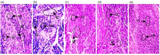
Figure 2(a-e) shows (a) Negative control showing normal histology of the spleen with the tissue parenchyma containing lymphocytes population, lymphatic nodule (LN), trabecula (TC), central arterioles (CA) and germinal centre (GM), (b) Positive control/lymphoid malignant group showing reactive spleen with the tissue parenchyma with lymphocyte population, enlarged trabeculae (TC) and mitotic bodies (MC) consistent with reactive spleen indicating rapid dividing cells, which is evidence of cancer and (c-e) showing 10, 20 and 30 mL kg–1 doses of body weight of Malayan green dwarf Hybridized immature coconut water groups respectively showing restoration of normal histology of the Spleen with the tissue parenchyma containing lymphocytes population(LC), lymphatic nodule (LN) and lymphoid follicular.
|
|
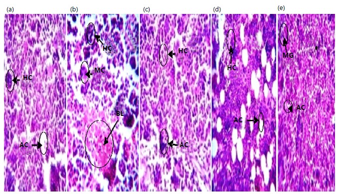
Figure 3(a-e) shows (a) Negative control showing normal histology of the bone marrow with hematopoietic cells (HC) and adipocytes (AC), (b) Positive control/lymphoid malignant group showing Reactive Bone marrow with hematopoietic cells (HC), invasion of bone marrow by blasts (BL) and mitotic bodies (MC) consistent with reactive bone marrow and (c-e) showing 10, 20 and 30 mL kg–1 doses of body weight of Malayan green dwarf Hybridized immature coconut water groups respectively showing restoration of normal histology of the Bone marrow with hematopoietic cells (HC), Megakaryocytes (MG) and adipocytes (AC).
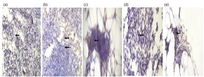
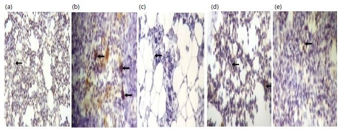
Figure 4(a-e) shows (a) Negative control reveals a negative nuclear immunohistochemistry reaction (black arrow), (b) Positive control/lymphoid malignant group reveal selective positive nuclear immunohistochemistry reaction (black arrow) and (c-e) showing 10, 20 and 30 mL kg–1 doses of body weight of Malayan green dwarf Hybridized immature coconut water groups respectively revealing restoration of normal immunohistochemistry of the inguinal lymph node with negative nuclear immunohistochemistry reaction (black arrow).
|
|
Figure 5(a-e) shows (a) Negative control reveals a negative cytoplasmic staining pattern (black arrow), (b) Positive control/lymphoid malignant group reveal a positive cytoplasmic staining pattern (black arrow) and (c-e) showing 10, 20 and 30 mL kg–1 doses of body weight of Malayan green dwarf Hybridized immature coconut water groups respectively revealing restoration of normal immunohistochemistry of the inguinal lymph node with negative cytoplasmic staining pattern (black arrow).
DISCUSSION
This study demonstrated the successful induction of lymphoid malignancy in Wistar rats intraperitoneal with 0.14 mL kg–1 b.wt., of benzene 48 hrs for 4 weeks as seen in the histology reports in the inguinal lymph node, spleen and bone marrow in the positive control group (benzene-induced) showing the cortex populated with mixed populations of lymphocytes in a lymph node, parenchyma with lymphocytes population, enlarged trabeculae in the spleen, hematopoietic cells, adipocytes, megakaryocytes, the invasion of bone marrow by the blasts and all the tissues displaying classic mitotic bodies and multiple nucleation’s consistent with reactive process indicating rapid dividing cells which is evidence of cancer with overexpression of p53 and Bcl-2 cancer biomarkers. Benzene-induced lymphoid malignancy is consistent with previous studies where benzene was used to induce leukaemia6,12. Overexpression of Bcl-2 is consistent with previous studies that found it to be a feature of lymphoid malignancy13,14 and determination of Bcl-2 expression by immunohistochemistry was reproducible and comparable to flow cytometry and immunoblotting15. Overexpression of p53 is in agreement with another study in which it was observed that p53 overexpression might be an indicator of cancer that is aggressive and poor prognosis16.
However, the histology reports of the inguinal lymph node, spleen and bone marrow in negative control show the cortex populated with lymphocytes, displaying memory cells and small non-cleaved cells consistent with normal histology of the lymph node as seen in the lymph node; the tissue parenchyma with lymphocytes population, lymphatic nodule, trabecule, central arterioles and germinal centre consistent with normal histology of the spleen as observed in spleen and haematopoietic cells, adipocytes as seen in bone marrow without expression of p53 and Bcl-2 cancer biomarkers.
Furthermore, treatment with 10, 20 and 30 mL kg–1 b.wt., of Malayan green dwarf hybridized immature (HI) coconut water: Restoration of normal histology of the lymph node with small non-cleaved cell (SNC), lymphoid follicle (LF), lymphocytes (LC), germinal centre (GC), fats cells (FC), perifollicular mantle (PM), memory cells (M) and post-capillary venule (PCV) restoration of normal histology of the Spleen with the tissue parenchyma containing lymphocytes population (LC), lymphatic nodule (LN) and lymphoid follicular; restoration of normal histology of the bone marrow with hematopoietic cells (HC), Megakaryocytes (MG) and adipocytes (AC), restoration of normal immunohistochemistry of the inguinal lymph node with negative nuclear immunohistochemistry reaction, restoration of normal immunohistochemistry of the inguinal lymph node with negative cytoplasmic staining pattern.
The phytotherapeutic effect observed in the spleen, bone marrow, lymph node histology and immunohistochemistry results in this study could be due to Malayan green dwarf hybridized immature coconut water bioactive phytochemicals such as Phenolic Compounds, Alkaloids, Saponins, Glycosides and Triterpenoids that possess anticancer properties.
Phenolic compounds express better anticancer activity based on the number of hydroxylic groups found in the compounds17.
Alkaloids, Saponins and Glycosides’ anticancer mechanism activities are by blocking cell proliferation by inducing cell cycle arrest at the G1 or G2/m phases18.
Triterpenoids block the initiation and promotion of carcinogenesis and create neoplasm cell differentiation and apoptosis19.
This finding was supported by the report of previous works conducted by various researchers in which exposure to benzene resulted in leukemia but was ameliorated by natural plant extracts that possess such phytochemical bioactive substances6,12,20.
This also was consistent with previous research showing that therapeutics targeting Bcl-2 and p53 have significant clinical activity in the treatment of lymphoid malignancy21,22. This also indicates the phytotherapeutic effect of Malayan green dwarf hybridized immature (HI) coconut water on benzene-induced lymphoid malignancy. The doses of 10, 20 and 30 mL kg–1 b.wt., of Malayan green dwarf hybridized immature (HI) coconut water for phytotherapeutic were observed to be more potent. This was consistent with a study on the Prophylactic Potential of Tender Coconut Water on Haematologic Disorder in Benzene-Induced Lymphoid Malignancy in Wister Rats where a dosage of 10 mL kg–1 b.wt., daily was reported6.
This study implies that Malayan green dwarf hybridized immature (HI) coconut water is potent anticancer therapy and can be applied in the management of lymphoid malignancy. However, the bioactive phytochemical substance could not be extracted separately, hence further work is recommended in this area.
CONCLUSION
This study reveals the phytotherapeutic effect of bioactive phytochemicals of Malayan green dwarf hybridized immature (HI) coconut water on benzene-inducing lymphoid malignancy. The discovered doses for such phytotherapeutic effects were 10, 20 and 30 mL kg–1 b.wt., daily of Malayan green dwarf hybridized immature (HI) coconut water. These doses will assist in clinical trials in human beings. Further work can be carried out to extract each component of the bioactive phytochemicals for novel anticancer therapy.
SIGNIFICANCE STATEMENT
Chemotherapy is commonly used for the treatment of lymphoid malignancy with associated serious side effects and complications with little or no improvement in patient survival and they are very expensive. Therefore, there is a need for immediate development of an effective therapy with little or no side effects and with a low cost. This study offers alternative natural therapy for lymphoid malignancy using Malayan green dwarf hybridized immature (HI) coconut water with no side effects at a low cost. This study will help the researcher uncover the critical area of lymphoid malignancy severity that many researchers were not able to explore. Thus, a new theory on this Malayan green dwarf hybridized immature (HI) coconut water on lymphoid malignancy may be arrived at.
ACKNOWLEDGMENTS
Our sincere appreciation to Prof. (Mrs). E.O. Osime in the Medical Laboratory Department, University of Benin, Edo State, Nigeria, who supervised the research work. We will not fail to acknowledge Dr. Oboma Yibala and Mr. Chukwuma Anekwe for their technical support.
REFERENCES
- Hanahan, D. and R.A. Weinberg, 2011. Hallmarks of cancer: The next generation. Cell, 144: 646-674.
- Rodriguez-Abreu, D., A. Bordoni and E. Zucca, 2007. Epidemiology of haematological malignancies. Ann. Oncol., 18: 13-18.
- Onuoha, E.C., M.E. Ike, T.A. Diepreye, E.F. Hallie, V.A. Maduka, N. Nelson-Ebimie and J.N. Nden, 2019. Haematological changes in patients with lymphoid malignancies on chemotherapy in Benin City, Edo State, Nigeria. J. Med. Physiol. Biophys., 61: 34-38.
- Tan, T.C., L.H. Cheng, R. Bhat, G. Rusul and A.M. Easa, 2014. Composition, physicochemical properties and thermal inactivation kinetics of polyphenol oxidase and peroxidase from coconut (Cocos nucifera) water obtained from immature, mature and overly-mature coconut. Food Chem., 142: 121-128.
- Rattan, S.I.S. and L. Sodagam, 2005. Gerontomodulatory and youth-preserving effects of zeatin on human skin fibroblasts undergoing aging in vitro. Rejuvenation Res., 8: 46-57.
- Elekwa, I., V.C. Ude, O. Emmanuel, V.O. Amachaghi and E.A. Ugbogu, 2021. In vivo studies on the ameliorative effect of coconut water against carbon tetrachloride induced toxicity in rats. Biomarkers, 26: 570-577.
- Snyder, R., 2012. Leukemia and benzene. Int. J. Environ. Res. Public Health, 9: 2875-2893.
- Vlaanderen, J., Q. Lan, H. Kromhout, N. Rothman and R. Vermeulen, 2012. Occupational benzene exposure and the risk of chronic myeloid leukemia: A meta-analysis of cohort studies incorporating study quality dimensions. Am. J. Ind. Med., 55: 779-785.
- Schnatter, A.R., D.C. Glass, G. Tang, R.D. Irons and L. Rushton, 2012. Myelodysplastic syndrome and benzene exposure among petroleum workers: An International Pooled Analysis. J. Natl. Cancer Inst., 104: 1724-1737.
- Wu, C.C., J.R. Blount, A. Haimbaugh, S. Heldman, J.N. Shields and T.R. Baker, 2022. Evaluating phenotypic and transcriptomic responses induced by low-level VOCs in zebrafish: Benzene as an example. Toxics, 10.
- Lorke, D., 1983. A new approach to practical acute toxicity testing. Arch. Toxicol., 54: 275-287.
- Olufemi, A.E., F. Ayodeji, A.R. Adekemi, B.E. Oluseyi, A.F. Ajoke, A.O. Aminat and L.O. Idris, 2017. African polyherbal formulation possesses chemopreventive and chemotherapeutic effects on benzene- induced leukemia in Wistar rats. Annu. Res. Rev. Biol., 16.
- Agarwal, B. and K.N. Naresh, 2002. Bcl-2 family of proteins in indolent B-cell non-Hodgkins, lymphoma: Study of 116 cases. Am. J. Hematol., 70: 278-282.
- Roberts, A.W., M.S. Davids, J.M. Pagel, B.S. Kahl and S.D. Puvvada et al., 2016. Targeting BCL2 with venetoclax in relapsed chronic lymphocytic leukemia. N. Engl. J. Med., 374: 311-322.
- Onel, B., M. Carver, G. Wu, D. Timonina, S. Kalarn, M. Larriva and D. Yang, 2016. A new G-quadruplex with hairpin loop immediately upstream of the human BCL2 P1 promoter modulates transcription. J. Am. Chem. Soc., 138: 2563-2570.
- Abdul-Razaq, H.K., M.H. Alhawaz and N.H.J. Al-Echrish, 2016. Evaluation of the p53 gene expression in breast cancer in respect to age, grade, stage and lymph nodes. Basrah J. Surg., 22: 17-25.
- Chen, M., H. Meng, Y. Zhao, F. Chen and S. Yu, 2015. Antioxidant and in vitro anticancer activities of phenolics isolated from sugar beet molasses. BMC Complementary Altern. Med., 15.
- Burgeiro, A., C. Gajate, E.L.H. Dakir, J.A. Villa-Pulgarín, P.J. Oliveira and F. Mollinedo, 2011. Involvement of mitochondrial and B-RAF/ERK signaling pathways in berberine-induced apoptosis in human melanoma cells. Anti-Cancer Drugs, 22: 507-518.
- Patlolla, J.M.R. and C.V. Rao, 2012. Triterpenoids for cancer prevention and treatment: Current status and future prospects. Curr. Pharm. Biotechnol., 13: 147-155.
- Maher, T., R.A. Raus, D. Daddiouaissa, F. Ahmad and N.S. Adzhar et al., 2021. Medicinal plants with anti-leukemic effects: A review. Molecules, 26.
- Demir, S., E. Boldrin, Q. Sun, S. Hampp and E. Tausch et al., 2020. Therapeutic targeting of mutant p53 in pediatric acute lymphoblastic leukemia. Haematologica, 105: 170-181.
- Kapoor, I., J. Bodo, B.T. Hill, E.D. Hsi and A. Almasan, 2020. Targeting BCL-2 in B-cell malignancies and overcoming therapeutic resistance. Cell Death Dis., 11.
How to Cite this paper?
APA-7 Style
Emmanuel,
O.C., Hallie,
E.F. (2023). Ameliorative Effect of Tender Coconut Water on Benzene Induced Lymphoid Malignancy in Wistar Rat. Asian Journal of Biological Sciences, 16(4), 493-501. https://doi.org/10.3923/ajbs.2023.493.501
ACS Style
Emmanuel,
O.C.; Hallie,
E.F. Ameliorative Effect of Tender Coconut Water on Benzene Induced Lymphoid Malignancy in Wistar Rat. Asian J. Biol. Sci 2023, 16, 493-501. https://doi.org/10.3923/ajbs.2023.493.501
AMA Style
Emmanuel
OC, Hallie
EF. Ameliorative Effect of Tender Coconut Water on Benzene Induced Lymphoid Malignancy in Wistar Rat. Asian Journal of Biological Sciences. 2023; 16(4): 493-501. https://doi.org/10.3923/ajbs.2023.493.501
Chicago/Turabian Style
Emmanuel, Onuoha, Chinedu, and Ezekiel Fayiah Hallie.
2023. "Ameliorative Effect of Tender Coconut Water on Benzene Induced Lymphoid Malignancy in Wistar Rat" Asian Journal of Biological Sciences 16, no. 4: 493-501. https://doi.org/10.3923/ajbs.2023.493.501

This work is licensed under a Creative Commons Attribution 4.0 International License.