Post-Hatch Age Related Development of Spleen of Broiler Chicken: A Biometric and Histomorphometric View
-
Shabnaz Aktar
Department of Veterinary and Animal Sciences, University of Rajshahi, Rajshahi 6205, Bangladesh
Mst. AeshaDepartment of Veterinary and Animal Sciences, University of Rajshahi, Rajshahi 6205, Bangladesh
Md. Monirul IslamDepartment of Veterinary and Animal Sciences, University of Rajshahi, Rajshahi 6205, Bangladesh
Md. Royhan Gofur

Department of Veterinary and Animal Sciences, University of Rajshahi, Rajshahi 6205, Bangladesh
| Received 13 Sep, 2023 |
Accepted 19 Nov, 2023 |
Published 31 Dec, 2023 |
Background and Objective: The primary organ of systemic immunity in birds is the spleen. In chickens, the spleen is the largest peripheral lymphoid organ, although little is known about how the spleen develops after hatching. Since the spleen plays a significant role in broiler chickens’ defensive mechanisms, the present study aimed to examine the age-related post-hatch development of the spleen, specifically its biometry and histomorphometry at various post-hatch developmental stages. Materials and Methods: The spleens of six different post-hatch developmental stages (post hatch day 1, day 7, day 14, day 21, day 28 and day 35) were taken after an ethical sacrifice (the cervical subluxation method) and subjected to biometric and histomorphometric evaluations. Biometric parameters (weight, length, width and thickness) were measured and for histomorphometrical study, selected formalin-fixed samples were processed and stained for microscopic analysis using a typical Mayer's Hematoxylin and Eosin stain. Results: The parenchyma of the spleen is formed of red pulp and white pulp that are intermingled with a lack of trabeculae and an identifiable marginal zone in all post-hatch age groups. A rising tendency in spleen capsule thickness, white pulp and lymphatic nodule length and breadth was noted. However, from day 14 onward, the white pulp size and the capsular thickness and from day 21 onward, the lymphatic nodules size were significantly (p<0.05) different among age groups. Conclusion: Although histologic structures were almost similar to other birds unlike mammals, values for the biometric and histomorphometric parameters were different from other bird species and even from indigenous and Sonali chicken.
| Copyright © 2023 Aktar et al. This is an open-access article distributed under the Creative Commons Attribution License, which permits unrestricted use, distribution, and reproduction in any medium, provided the original work is properly cited. |
INTRODUCTION
Designing a poultry health management program requires a thorough understanding of the avian immune system. The mechanisms of immunosuppression and the creation of methods to improve immunological response in commercial poultry are currently of great interest1. To fully comprehend the physiology and immunology of lymphoid tissue, one must first have a general understanding of its composition. Similar to mammals, the major lymphoid organs and secondary lymphoid organs are the two different morphological and functional elements of the avian lymphoid system2. The existence of the bursa of Fabricius, which is comparable to bone marrow in mammals and the thymus as principal lymphoid organs distinguish the immune system (lymphoid) of birds most significantly from that of mammals. Having no lymphnodes, the bone marrow, Gut-Associated Lymphoid Tissue (GALT), Mucosa-Associated Lymphoid Tissue (MALT) and spleen are the secondary lymphoid organs3.
Bird immune system strength has been inferred from studies of avian ecology, parasitology and evolution using the avian spleen4. The spleen performs a number of advantageous functions in immune response, including purging the bloodstream of infected or damaged cells and giving the host immunity to infection5. The chicken immune system is crucial in phylogeny because it lacks lymph nodes but contains the bursa of Fabricius6. Avians and mammals have different immune systems in terms of their anatomical makeup. Although the mammalian spleen parenchyma consists of red and white pulp with a distinct marginal zone, the chicken spleen lacks a morphologically defined marginal zone7. Compared to its mammalian equivalent, the white pulp’s architecture is much different. There are three morphologically different regions in the chicken spleen: (1) Periarteriolar lymphocyte sheaths (PALS) that surround arterioles, (2) Peri- ellipsoid lymphocyte sheaths (PELS) which is the lymphoid tissue of the B lymphocytes that surround penicillary capillaries and (3) Follicles with germinal centers (the primary site of B lymphocytes proliferation and differentiations), which are surrounded by a capsule of connective tissue. However, the fact that a corona and marginal zone are morphologically indistinguishable, however, suggests that the PELS is the chicken equivalent of the mammalian marginal zone7. The white pulp of chicken spleens is well known to have a crucial role in the first immune response, particularly in defense against blood-borne antigens5,8.
With sexual maturity, the development of the peripheral and central immune organs slow and eventually stop9. Immune function maintenance is tightly linked to the development of the peripheral lymphoid organs10. In infectious disorders, the size of the spleen is utilized as a measure of immune system response11. Chickens’ spleen size dynamics show that as they grow, the mass of their spleen increases in direct proportion to their body masses. Although extensive research has already been done on the biometry and histomorphometry of the post-hatch developing spleen in Deshi and Sonali chicken, broiler chicken literature was not readily available despite its importance to the poultry industry as well as the national economy in Bangladesh. The current study was designed using a biometric and histomorphometric approach to analyze the spleen of broiler chickens at various post-hatch developmental phases.
MATERIALS AND METHODS
Animal experiments were implemented according to the guidelines set by the Institutional Animal, Medical Ethics, Biosafety and Biosecurity Committee of the University of Rajshahi, Bangladesh. A total of 120 day-old broiler chicks (EP, Efficiency Plus) were procured from Aftab Bahumukhi Farms Limited, Bangladesh. The day-old chicks had normal bodily functions and physical development. The study was carried out from January, 2023 to August, 2023. The experimental broiler chickens were reared in Narikel Bariya Poultry Farm run by the Department of Veterinary and Animal Sciences, University of Rajshahi, Bangladesh. The brooding time of chicks was 7 days. The experimental chickens were raised in hygienic conditions with unlimited access to food and water. The experimental chicken was reared feeding the broiler starter (days 1 to 14), broiler grower (days 15 to 28) and broiler finisher (days 29 to 36) purchased from Nourish Feeds Limited, Bangladesh. The immunization schedule was followed. The homogeneity of the management practice was preserved as much as feasible over the entire experimental period. The poultry shed had adequate airflow and ventilation. Both the farm and the poultry shed’s biosecurity were strictly maintained. The chickens that were gathered did not exhibit any developmental issues or diseases that could have interfered with the experiment or affected its outcome.
Post-hatch developmental age groups: The experimental chickens were divided into six age groups, post-hatch day 1, day 7, day 14, day 21, day 28 and day 35, having twenty chickens in each group.
Tissue collection and preservation: Chickens used in the experiment were slaughtered using the cervical subluxation technique on days 1, 7, 14, 21, 28 and 35. After the birds were sacrificed, the spleen samples were taken by dissection and cleansed in physiological saline. (0.9%). After biometric analysis, the collected samples were immediately placed in a 10% formalin solution for further histomorphometric study.
Biometric study: Gross morphometric (weight, length, width and thickness) values of the spleen of different age groups of broiler chickens were examined. The spleen’s weight was measured using an electronic digital weighing scale (Model: PS.P3.310, Taiwan) in grams (gm) and the organ’s size was determined using slide callipers on a millimeter (mm) scale.
Histomorphometrical study: The selected formalin-fixed samples were processed in the laboratory according to the usual histological method for paraffin sections. Sections were cut at a thickness of 5 m and stained for microscopic analysis using a typical Mayer’s Hematoxylin and Eosin (H & E) stain, with certain modifications, according to Gofur et al.12. The stained sections of the post-hatch developing spleen were carefully investigated using compound microscopes at magnifications of 10 and 40. The images of the stained tissue sections were taken using a photographic microscope system (digital camera model: C-B5, OPTIKA, Italy equipped with a microscope, Model B-293PLi, OPTIKA, Italy). A pre-calibrated ocular micrometer (Erma, Japan) in μm was used to measure the histomorphometric characteristics (the length and breadth of the white pulp and the thickness of the capsule).
Statistical analyses: All observations were represented as mean±SE. Differences in biometrical and histomorphometrical values of spleen among the different post-hatch developing ages of broiler chickens were evaluated by One-way Analysis of Variance (ANOVA), followed by Turkey HSD post hoc analysis according to Gofur et al.13. Significant differences were defined as p values of 0.05 or less.
RESULTS
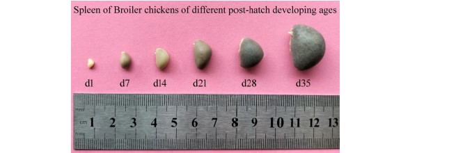
Biometry of the spleen in post-hatch developing broiler chickens: The biometrical parameters (weight, length, width and thickness) of the spleen of broiler chickens were measured and observed that post-hatch age had an influence on the growth of the spleen of broiler chickens. The biometric values of the spleen were increased with the advancement of post-hatch developing age (Table 1 and Fig. 1). It was observed that the biometric values of the spleen were significantly and gradually increased in every advanced post-hatch age group throughout the whole experimental period except the weight of spleen that was significantly differed from post-hatch day 21 in broiler chickens. Compared to day 1, the splenic length, width and breadth expanded by 4 to 5 times and the weight increased by around 50 times at day 35 of post-hatch age.
|
|
| Table 1: | Biometrical values of broiler spleen at different post-hatch developing ages | |||
| Age groups | Weight (g) |
Length (mm) |
Width (mm) |
Thickness (mm) |
| Day 1 | 0.045±0.006a |
5.10±0.10a |
3.80±0.10a |
2.47±0.13a |
| Day 7 | 0.187±0.011a |
7.93±0.47b |
5.37±0.28b |
4.87±0.18b |
| Day 14 | 0.299±0.012ab |
12.33±0.79c |
8.03±0.28c |
6.97±0.35c |
| Day 21 | 0.833±0.058b |
15.17±0.39d |
11.83±0.15d |
8.93±0.32d |
| Day 28 | 1.458±0.132c |
18.17±0.39e |
13.33±0.32e |
10.67±0.39e |
| Day 35 | 2.318±0.215d |
22.07±0.57f |
16.17±0.39f |
12.57±0.40f |
| Mean±SE and values with several superscript alphabets within the same column signify a significant age difference (p<0.05) | ||||
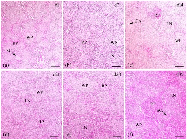
Histomorphometry of the spleen in post-hatch developing broiler chickens: A thin capsule made of collagen and smooth muscle fibers encircled the histological architecture of the spleen on post-hatch day 1 in broiler chickens. There were no splenic trabeculae. White pulp with blood vessels and red pulp made up the splenic parenchyma. The red pulp stood out among the white pulps and was dispersed widely. Like in mammals, there was no discernible marginal zone between the pulps. Sheathed capillaries were observed at the white pulp (Fig. 2a). Broiler chicken spleens were examined on post-hatch day 7 and found to be identical to those examined on post-hatch day 1 (Fig. 2b). The parenchyma of the spleen contained a prominent lymphatic nodule at this developmental stage (Fig. 2b). The thick splenic capsule was present without any trabeculae on day 14 of post-hatch development. The red pulp was distinct and it was dispersed among the white pulp. At this point, the central artery was identified. Distinct lymphatic nodule was also observed (Fig. 2c). At days 21, 28 and 35 of post-hatch development, the histological characteristics of the spleen in broiler chickens were identical to those at day 14, with histological structures more advanced than the earlier groups. Distinct lymphatic nodule and sheathed capillary were observed at white pulp at every stages of post-hatch development (Fig. 2d-f).

|

|
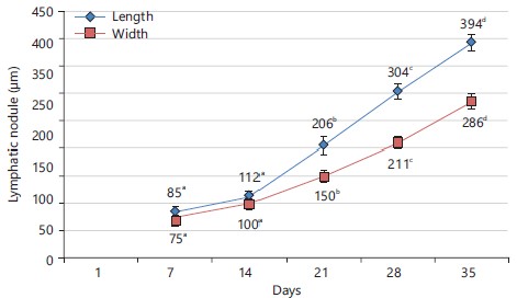
During the post-hatch development of the broiler spleen, the variations in the spleen capsule’s thickness, the length and width of white pulp and length and breadth of lymphatic nodules in different post-hatch age groups were shown in Fig. 3, 4 and 5, respectively. An increasing trend in the thickness of capsule of the spleen, length and breadth of white pulp and length and breadth of lymphatic nodules were noticed throughout the experimental period, until post-hatch day 35 in broiler chicken. However, thickness of capsule and length and breadth of white pulp significantly (p<0.05) differ among post-hatch age groups from day 14 onwards, whereas length and breadth of lymphatic nodules significantly (p<0.05) differ from day 21 onwards. Compared to day 1, the thickness of the splenic capsule expanded by 3 times and the length and breadth of white pulp expanded by around 5 times on day 35 of post-hatch age, whereas the length and breadth of lymphatic nodules expanded by around 4-5 times on day 35 compared to day 14 when the lymphatic nodules were first appeared in splenic parenchyma.
|
DISCUSSION
The size of the avian spleen, an important immunological organ in birds, can be employed as an indicator of the immune system’s responses under various conditions. It was observed that the biometric values of the spleen were significantly and gradually increased in every advanced post-hatch age group throughout the whole experimental period except the weight of the spleen was significantly different from post-hatch day 21 in broiler chickens. The immune system begins to grow and develop in broiler chickens early in life14. Spleen weight may increase quickly in the first few weeks of life as a result of the splenic parenchyma's histological maturation phenomenon15. The spleen of broiler chicken in all postnatal ages had a thin outer fibromuscular capsule covering it, no trabeculae extend into the parenchyma. The parenchyma was composed of both white and red pulp, with no boundary (marginal zone) between them. However, broiler chicken has certain distinctive characteristics. On hatching day, there were no lymphatic nodules inside the splenic parenchyma. As broilers age, their lymphocytes start to group together and by day 7, there were clear lymphatic nodules that were more developed and bigger with age. In the present research the histological thickness of the capsule, length and breadth of white pulp and length and breadth of lymphatic nodules showed a significantly increasing trend until post-hatch day 35 in broiler chicken.
The biometrical parameters of the spleen in broiler chickens were considerably rising up to day 35, showing developmental progress which is consistent with the findings in Sonali chickens where Ayman et al.16 stated that up until day 56, the spleen’s weight, length, width and thickness were all noticeably expanding. The biometric values of the spleen were found to be increasing with the increase of post-hatch age in Deshi chicken reported by Khalil et al.17 and in White Pekin duck reported by Indu et al.18. According to research, the spleen in chickens reaches its greatest size in the first six weeks after birth19. Hodges20 reported that the spleen's size and weight varies with age in various breeds and even within the same breed under various circumstances.
Broiler chicken spleens were found to have a comparable general histological structure to that of other chicken breeds reported in Sonali chicken16, in Deshi chicken17, also in indigenous ducklings of Bangladesh21, in ostrich22 and in Japanese quail23. Zhang et al.5 reported a similar architecture of the spleen in adult broiler chickens in general. The trabeculae were poorly developed in other avian species like in ostrich, a non-flying bird (Struthio camelus) spleen22, Japanese quail (Coturnix coturnix Japonica) spleen23, Eurasian moorhen (Gallinula chloropus, also known as the “swamp chicken”) spleen24 and Red-Legged Partridge (Alectoris chukar) spleen25. A significantly increasing trend in histomorphometrical parameters of capsule, white pulp and lymphatic nodules was observed in broiler chicken until post-hatch day 35. Previously similar significantly increasing trend was reported in Sonali chicken from day 1 to day 5616. However, the thickness of the capsule was a little higher in broiler chicken (51.80±2.15 μm) at post-hatch day 35 than that of Sonali chicken (32.34±5.754 μm at day 56)16. Although the presence of lymphatic nodules in the splenic parenchyma has previously been recognized, their histomorphometric values have not been determined16,17. Ayman et al.16 reported lymphatic nodules appeared in splenic parenchyma from day 14 onwards in Sonali chickens, whereas lymphatic nodules were observed from day 7 onwards in broiler chickens indicating that breed diversity may affect when lymphatic nodules first occur. However, Song et al.2 concluded that the broiler chicken immune system and its function are not fully formed until day 13 and the immune system does not reach maturity until day 34 in broiler chickens. In the present study, the samples were collected a weekly intervals. It will be better to collect the tissue sample daily or every alternative day for a detailed and in-depth microscopic study of the broiler spleen. Basic knowledge of the post-hatch age-related development of the broiler chicken spleen from day 1 to day 35 will surely advance understanding and create a wealth of new chances for further immunological research and also aid in the diagnosis of different poultry diseases.
CONCLUSION
The morphologic features of the spleen in broiler chickens were different from other bird species and even from indigenous and Sonali chicken, but histologic structures were almost similar to indigenous and Sonali chicken, duck and quail spleens. The broiler spleen parenchyma was formed of red pulp and white pulp that is intermingled with lacked trabeculae and an identifiable marginal zone in all post-hatch age groups. Throughout the investigation, a substantial relationship between post-hatch age and splenic development was noted. An increasing trend in values of the biometric and histomorphometric parameters of the broiler spleen was observed. Further study is needed to understand the ultrastructure of the boiler spleen and to provide in-depth clarification of the role of the spleen in broiler immunity.
SIGNIFICANCE STATEMENT
The rising poultry sector contributed a lot to raising the national economy in Bangladesh. Any disorder affecting the immune organs results in immunosuppression that leads to an increased rate of mortality in broiler chickens. The present study depicted the gradual gross and microscopic development of broiler spleen from hatching day to post-hatch day 35 and established a significant correlation between post-hatch age and splenic development. Impairment or deviation of normal splenic development indicates that the broiler suffers from any disorder or disease. Moreover, the results of the present post-hatch age-related study open up a wide range of possibilities for future molecular studies and aid in the detection of many disorders affecting poultry.
ACKNOWLEDGMENT
The Ministry of Science and Technology of Bangladesh provides financial support for the research (special allocation Project ID: SRG-221300).
REFERENCES
- Li, C., L. Wang and S. Zheng, 2023. Editorial: Immunosuppressive disease in poultry. Front. Immunol., 14.
- Song, B., D. Tang, S. Yan, H. Fan and G. Li et al., 2021. Effects of age on immune function in broiler chickens. J. Anim. Sci. Biotechnol., 12.
- Casteleyn, C., M. Doom, E. Lambrechts, W. van den Broeck, P. Simoens and P. Cornillie, 2010. Locations of gut-associated lymphoid tissue in the 3-month-old chicken: A review. Avian Pathol., 39: 143-150.
- Smith, K.G. and J.L. Hunt, 2004. On the use of spleen mass as a measure of avian immune system strength. Oecologia, 138: 28-31.
- Zhang, Q., Y. Waqas, P. Yang, X. Sun and Y. Liu et al., 2017. Cytological study on the regulation of lymphocyte homing in the chicken spleen during LPS stimulation. Oncotarget, 8: 7405-7419.
- Lowenthal, J.W., A.G.D. Bean and M.H. Kogut, 2013. What’s so special about chicken immunology? Dev. Comp. Immunol., 41: 307-309.
- Zhang, Q., X. Sun, T. Wang, B. Chen, Y. Huang, H. Chen and Q. Chen, 2019. The postembryonic development of the immunological barrier in the chicken spleens. J. Immunol. Res., 2019.
- Mebius, R.E. and G. Kraal, 2005. Structure and function of the spleen. Nat. Rev. Immunol., 5: 606-616.
- Gordon, J. and N.R. Manley, 2011. Mechanisms of thymus organogenesis and morphogenesis. Development, 138: 3865-3878.
- Nagy, N. and I. Bódi and I. Oláh, 2016. Avian dendritic cells: Phenotype and ontogeny in lymphoid organs. Dev. Comp. Immunol., 58: 47-59.
- John, J.L., 1994. The avian spleen: A neglected organ. Q. Rev. Biol., 69: 327-351.
- Gofur, M.R., M.Z.I. Khan, M.R. Karim and M.N. Islam, 2008. Histomorphology and histochemistry of testis of indigenous bull (Bos indicus). Bangladesh J. Vet. Med., 6: 67-74.
- Gofur, M., M. Sadi, S. Aktar, A. Khatun and M. Awal et al., 2023. Biometrical and histomorphometrical changes of testis in the dynamics of postnatal ontogenesis from birth to puberty of black Bengal goat. J. Adv. Vet. Anim. Res., 10: 237-243.
- Russell, A.R.B. and S.H. Murch, 2006. Could peripartum antibiotics have delayed health consequences for the infant? BJOG: Int. J. Obstet. Gynaecol., 113: 758-765.
- Olah, I. and B. Glick, 1982. Splenic white pulp and associated vascular channels in chicken spleen. Am. J. Anat., 165: 445-480.
- Ayman, U., M.R. Alam and S.K. Das, 2021. The spleen of sonali chicken: Morphohistology and biometric analysis at post hatching ages. Asian J. Med. Biol. Res., 7: 69-75.
- Khalil, M., S.Z. Sultana, M. Rahman, S. Mannan and S. Ahmed et al., 2009. Study of prenatal and postnatal development of spleen of Gallus domesticus (deshi chicken). Mymensingh Med. J., 18: 169-174.
- Indu, V.R., J.J. Chungath, K.R. Harshan, K.M. Lucy and S. Maya, 2000. Postnatal development of spleen in the White Pekin duck. Indian J. Poult. Sci., 35: 32-34.
- Chen, L.T., 1978. Microcirculation of the spleen: An open or closed circulation? Science, 201: 157-159.
- Hodges, R.D., 1974. The Histology of the Fowl. Academic Press, Cambridge, Massachusetts, ISBN: 9780123513502, Pages: 648.
- Sultana, N., M.Z.I. Khan, M.A. Wares and M.A. Masum, 2011. Histomorphological study of the major lymphoid tissues in indigenous ducklings of Bangladesh. Bangladesh J. Vet. Med., 9: 53-58.
- Kozlu, T., E.K. Sari, Y.A. Bozkurt and H. Altunay, 2011. A comparative study on the histological structure of the spleen in the ostrich (Struthio camelus), the kestrel (Falco tinnunculus) and the osprey (Pandion haliaetus). Acta Biol. Hung., 62: 113-121.
- Mustafa, F.E.Z.A. and S.M.M. El-Desoky, 2020. Architecture and cellular composition of the spleen in the Japanese quail (Coturnix japonica). Microsc. Microanal., 26: 589-598.
- Abdellatif, A.M., 2021. Structure of the Eurasian moorhen spleen: A comprehensive study using gross anatomy, light, and transmission electron microscopy. Microsc. Res. Tech., 84: 1696-1709.
- Özüdoğru, Z., H. Balkaya, H. Kara, A. Kara and D. Özdemir, 2021. Investigation of morphological and histological structure of red-legged partridge (Alectoris chukar) Spleen. Vet. Sci. Pract., 16: 57-62.
How to Cite this paper?
APA-7 Style
Aktar,
S., Aesha,
M., Islam,
M.M., Gofur,
M.R. (2023). Post-Hatch Age Related Development of Spleen of Broiler Chicken: A Biometric and Histomorphometric View. Asian Journal of Biological Sciences, 16(4), 514-521. https://doi.org/10.3923/ajbs.2023.514.521
ACS Style
Aktar,
S.; Aesha,
M.; Islam,
M.M.; Gofur,
M.R. Post-Hatch Age Related Development of Spleen of Broiler Chicken: A Biometric and Histomorphometric View. Asian J. Biol. Sci 2023, 16, 514-521. https://doi.org/10.3923/ajbs.2023.514.521
AMA Style
Aktar
S, Aesha
M, Islam
MM, Gofur
MR. Post-Hatch Age Related Development of Spleen of Broiler Chicken: A Biometric and Histomorphometric View. Asian Journal of Biological Sciences. 2023; 16(4): 514-521. https://doi.org/10.3923/ajbs.2023.514.521
Chicago/Turabian Style
Aktar, Shabnaz, Mst. Aesha, Md. Monirul Islam, and Md. Royhan Gofur.
2023. "Post-Hatch Age Related Development of Spleen of Broiler Chicken: A Biometric and Histomorphometric View" Asian Journal of Biological Sciences 16, no. 4: 514-521. https://doi.org/10.3923/ajbs.2023.514.521

This work is licensed under a Creative Commons Attribution 4.0 International License.