Uvaria chamae Stem Extracts Possess Nephroprotective and Cardioprotective Effects Against Carbon Tetrachloride-Induced Toxicity in Albino Rats
-
Jennifer Nkeiru Obas
Department of Biochemistry, Ebonyi State University, Abakaliki, Ebonyi 481101, Nigeria
David Chukwu Obasi

Department of Medical Biochemistry, David Umahi Federal University of Health Sciences, Uburu, Ebonyi, Nigeria
Kingsley Nwonu AgbafoDepartment of Biochemistry, Ebonyi State University, Abakaliki, Ebonyi 481101, Nigeria
| Received 29 Sep, 2023 |
Accepted 28 Nov, 2023 |
Published 31 Dec, 2023 |
Background and Objective: Renal and cardiovascular diseases are among the leading causes of death worldwide and require alternative medical interventions. This study investigated the effects of deionized water and ethyl acetate extracts of Uvaria chamae stem (DEEUS) on renal and cardiovascular parameters of carbon tetrachloride (CCl4)-induced toxicity in albino rats. Materials and Methods: A total of 54 male albino rats were divided into 9 groups of 6 rats each. Group 1 served as normal control. Groups 2-9 were injected with CCl4. Group 2 was untreated, group 3 received 20 mg kg-1 b.wt., of vitamin C, while groups 4-9 were treated with DEEUS at doses of 200, 400 and 600 mg kg-1 b.wt., respectively for 21 days via oral intubation. The renal and cardiovascular parameters of these rats were determined. The data obtained from the biochemical tests were subjected to a One-way Analysis of Variance (ANOVA). Results: There were significantly (p<0.05) higher concentrations of serum urea, creatinine, uric acid, sodium, potassium, bicarbonate, cTnI, TG, TC and LDL, with lower concentrations of HDL, as well as significant (p<0.05) higher activities of CK and LDH in the untreated group compared to the normal control. Upon treatment, the extracts prevented further tissue damage, leading to the recovery of the animals as shown by the differences between the treated and untreated groups. The effects were found to be dose-dependent, with higher effects observed in the ethyl acetate extract than in the deionized water extract. Conclusion: The results indicated that Uvaria chamae stem extracts possess nephroprotective and cardioprotective activities and could be harnessed for the management of renal and cardiovascular diseases.
| Copyright © 2023 Obas et al. This is an open-access article distributed under the Creative Commons Attribution License, which permits unrestricted use, distribution, and reproduction in any medium, provided the original work is properly cited. |
INTRODUCTION
Nephrotoxicity refers to the adverse effect of substances on renal function1. The function of kidney involves the excretion of water, solutes and metabolic wastes to maintain a constant internal environment (homeostasis) despite fluctuations in diet and fluid balance. The integrity of cells is dependent on the osmolality of the Extracellular Fluid (ECF), which is kept under tight control through osmoreceptors and volume receptors that allow the brain and kidneys to produce hormones and vasoactive substances to regulate salt and water excretion2. Measurement of Glomerular Filtration Rate (GFR) is used in the determination of kidney function. The GFR differs among species and pathological states of efferent and afferent arterioles such as vasoconstriction caused by drugs, which may affect GFR. The glomerular ultrafiltrate is produced when plasma is filtered through the glomerulus3. A key role of the kidney involves mediating the toxicity of natural substances, pollutants and many drugs. Some nephrotoxic drugs include antibiotics, cancer therapeutics, drugs of abuse and radiocontrast agents4. Nephrotoxicity causes alteration of kidney functions, resulting in kidney diseases with detrimental consequences.
Cardiovascular Diseases (CVD) are diseases of the heart and blood vessels and include cardiomyopathy, Coronary Heart Disease (CHD), Peripheral Arterial Disease (PAD), Rheumatic Heart Disease (RHD), Congestive Heart Failure (CHF), kinds of angina, stroke and myocardial infarction (MI), leading to mortality in humans globally5,6. The cause of MI is extensive myocardial tissue damage, developing as a result of discrepancies between oxygen demand and cardiomyocytes’ blood supply, regardless of atherosclerotic plaque7. Endocardial lipid peroxidation is caused by excess of reactive oxygen species generation, leading to the loss of cardioprotective antioxidants, increased oxidative stress and apoptosis7.
Carbon Tetrachloride (CCl4) is a chemical compound used for inducing toxicity through the production of free radicals in many tissues such as the heart, liver, kidneys, testis, lung, brain and blood8. The hepatic microsomal cytochrome P450 converts CCl4 to trichloromethyl-free radical9, which leads to the initiation of the lipid peroxidation process10. The formation of free radicals is the major mechanism of CCl4-induced cardiotoxicity, which is a rate-limiting process in tissue peroxidative damage11,12. This free radical and related reactive species may cause oxidative stress, which produces major interrelated rearrangements of cellular metabolism, an increase in intracellular free calcium, damage to membrane ion transport and permeability and destruction of the cells by lipid peroxidation13. This leads to loss of myocardial structural integrity and depressed cardiac function resulting in cardiotoxicity and congestive cardiac failure11,12.
Uvaria chamae is a plant of the Annonaceae family14,15. It is found mostly in the savanna and rainforest regions of Nigeria and some other African countries. It is called “Mmimiohia”, “Kaskaifi” and “Akisan” amongst the Ibos, Hausas and Yorubas, respectively16. The ripe fruits of U. chamae are yellow, with a sweet-edible pulp. The fruit carpels appear in finger-like clusters. The stem barks, root barks and leaves have various medicinal uses. A decoction of the stem is used in the treatment of diarrhea in Nigeria17. A study has shown that U. chamae possesses antioxidant and free radical scavenging potentials18. Pharmaceutically, chemical constituents of the leaves and bark of U. chamae have antifungal, antimalarial and bacteriostatic, especially cytostatic activities19. Methanol extract of U. chamae has been reported to neutralize some biological effects of Naja nigricollis venom20. The medicinal properties of U. chamae are attributed to the different phytochemicals it contains, such as alkaloids, flavonoids, saponins, phenols, tannins, steroids and glycosides as were observed at various concentrations in different solvent extracts15.
While synthetic drugs have been proven helpful in the treatment of various diseases, their use is limited due to their side effects21 and high costs which are not affordable in some rural areas. Hence, the need for medicinal plants. Based on the medicinal benefits of U. chamae and little scientific information on its nephroprotective and cardioprotective potentials against CCl4-induced toxicity, this study aimed to determine the cardioprotective effects of deionized water and ethylacetate extracts of U.chamae stem on Carbon Tetrachloride (CCl4)-induced toxicity in albino rats.
MATERIALS AND METHODS
Study area: This study was carried out at the Department of Biochemistry Research Laboratory, Faculty of Science, Ebonyi State University, Abakaliki, Nigeria, with approval from the Office of Research, Innovation and Institutional Ethics Committee (EBSU/BCH/ET/22/010). The study was started from November 2022 to March, 2023.
Reagents: Magnesium Chloride (MgCl2) solution, Sodium Hydroxide (NaOH), chloroform, Sodium Chloride (NaCl), distilled water, urea, creatinine, uric acid, sodium, potassium and Bicarbonate (HCO3–), Cardiac Troponin 1 (cTn1), creatine kinase, lactate dehydrogenase, cholesterol, triglycerides, high-density lipoprotein and low-density lipoprotein kits (Randox Laboratories Limited, BT29 4QY, United Kingdom) were used.
Equipments: The UV-Vis Spectrophotometer (Model 752, LKB, Bechion), refrigerator (Haier Thermocol, HTF-429H, England), Rotary Evaporator (Model 349/2 Corning Ltd., England), centrifuge (Haematospin 1400, Hawskey England), water bath (Julabo), weighing balance (Merek, England).
Collection and preparation of plant sample: Twenty fresh stems of Uvaria chamae were collected from Umuka-Okposi, Ohaozara Local Government Area, Ebonyi State, Nigeria. The stems were cut into small pieces and air-dried in the laboratory at room temperature with adequate turning to avoid fungal growth for 2 weeks. The dried stems were pulverized, sieved and stored in an air-tight plastic container. Two measures of powdered U. chamae stem of 500 g each were soaked in 2500 mL of deionized water and 2500 mL of ethyl acetate respectively, stirred, covered in an air-tight container and left to stand for 48 hrs. Afterwards, the extracts were filtered through a muslin cloth. The resulting deionized water (DW) and ethyl acetate (EA) extracts were concentrated and evaporated to dryness using a rotary evaporator (Model 349/2 Corning Ltd., England) at an optimum temperature of 50°C. The concentrated DW (51.40 g) and EA (54.60 g) extracts of U. chamae stem were stored in air-tight containers for analysis.
Animal management: One hundred adult male albino rats (weighing between 190 and 200 g) were purchased from animal house of Zoology Department, University of Nigeria, Nsukka and acclimatized for one week prior to the commencement of the experiment. They were kept at room temperature and maintained ad libitum on water and growers mash (feed).
Acute toxicity studies: An acute toxicity study, LD50, was performed according to Lorke method22. There were 11 groups (A to K) with 3 rats each. The rats were fasted overnight prior to the administration of the extracts. Afterwards, the animals in groups B to F and G to J received serial oral doses of 500, 1000, 2000, 4000 and 5000 mg kg–1 b.wt., of DW and EA extracts of U. chamae stem, respectively. Group A served as the normal control. After the administration of the extracts, food was withheld for a further 4 hrs. Animals were observed and monitored closely for signs of toxicity and lethality, individually for the first 30 min after dosing and periodically during the first 24 hrs and daily thereafter for 3 days. However, there was no death recorded, although physical signs of weakness (such as sluggish movement, poor eating and drinking) were observed in the groups given 4000 and 5000 mg kg–1 b.wt., after 3 days. Hence, 3 doses of 200, 400 and 600 mg kg–1 b.wt., of the extracts were chosen for the sub-acute toxicity study.
Preparations of drugs and treatment
Carbon Tetrachloride (CCl4): Prepared by mixing 10 mL of CCl4 and 10 mL of olive oil (1:1) and administered to the rats intraperitoneal at 2.5 mL kg–1 b.wt.
Deionized water and ethyl acetate extracts of U. chamae stem: Ten grams of each plant extract was separately dissolved in 50 mL of normal saline (1:5 w:v) as stock solution and was administered at 3 doses of 200, 400 and 600 mg kg–1 b.wt., respectively via oral intubation.
Vitamin C: One gram was dissolved in 50 mL of normal saline and administered to the rats at 20 mg kg–1 b.wt., via oral intubation.
Experimental design: Toxicity with 2.5 mL kg–1 b.wt., of CCl4 was induced in the animals and left for 1 hr before treatment with the plant extracts for 21 days via oral intubation. A total of 54 male albino rats were divided into 9 groups of six rats each as follows:
| • | Group 1: Normal control rats | |
| • | Group 2: CCl4 control rats-administered with CCl4 without treatment | |
| • | Group 3: Standard control rats-administered with CCl4 and treated with 20 mg kg–1 b.wt., of vitamin C | |
| • | Group 4: Administered with CCl4 and treated with 200 mg kg–1 b.wt., of deionized water extract (DWE) | |
| • | Group 5: Administered with CCl4 and treated with 400 mg kg–1 b.wt., of DWE | |
| • | Group 6: Administered with CCl4 and treated with 600 mg kg–1 b.wt., of DWE | |
| • | Group 7: Administered with CCl4 and treated with 200 mg kg–1 b.wt., of ethyl acetate extract (EAE) | |
| • | Group 8: Administered with CCl4 and treated with 400 mg kg–1 b.wt., of EAE | |
| • | Group 9: Administered with CCl4 and treated with 600 mg kg–1 b.wt., of EAE |
Blood samples were collected from five animals in each group after fasting for 24 hrs from the last treatment via ocular and cardiac punctures in anticoagulant-free bottles and centrifuged at 2000 rpm for 15 min to get the serum used for biochemical studies. Also, the kidneys and hearts of the rats were excised, rinsed, cleaned in normal saline and preserved in 10% formalin for Histopathological assessments.
Biochemical analyses
Kidney function parameters: Serum concentrations of urea and creatinine23, uric acid24, sodium and potassium25 and bicarbonate ion26 were carried out with reagent kits.
Cardiovascular parameters: Cardiac Troponin 1 (cTn1) concentration27, creatine kinase (CK) activity28, Lactate Dehydrogenase (LDH) activity29, total cholesterol concentration30, triglyceride (TG) concentration31, High-Density Lipoprotein Cholesterol (HDL) concentration30, Low-Density Lipoprotein Cholesterol (LDL) concentration32.
Histological examination of tissues: The experimental rats were humanely sacrificed at the end of the study. Sections of the kidneys and heart from each group were collected for histological examination. The fixing of the samples was done for 48 hrs with formalin buffered with 10% phosphate. Trimming and dehydration of the samples were in 4 alcohol grades (70, 80, 90% and absolute alcohol), after which they were cleared in 3 xylene grades and enclosed in molten wax. The blocks were divided into sections of 5 μm thick with a rotary microtome on solidifying and floated in a water bath, incubated at 60°C for 30 min. The tissue sections were further cleared in 3 xylene grades and rehydrated in 90, 80 and 70% alcohol grades. Then, hematoxylin was used to stain them for 15 min, before blueing with ammonium chloride. Afterwards, 1% acid alcohol was used for differentiation before counterstaining with Eosin. A mountant, DPX was used to make permanent mounts on degreased glass slides. Motic™ compound light microscope was used for examination of the slides with ×4, ×10 and ×40 objective lenses. A Motic™ 5.0 megapixel microscope camera was used to take the photomicrographs of the tissues at magnifications of ×160 and ×400.
Statistical analysis: The data obtained from the biochemical tests were subjected to a One-way Analysis of Variance (ANOVA). Significant differences were obtained at p<0.05 and the results were expressed as Mean±Standard Error of mean (SEM). The SPSS version 23 and Microsoft Excel 2016 software were used.
RESULTS
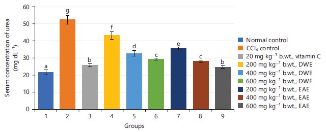
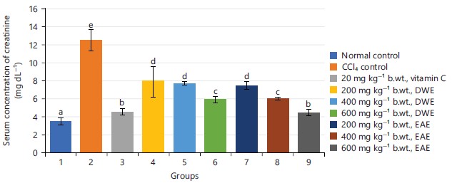
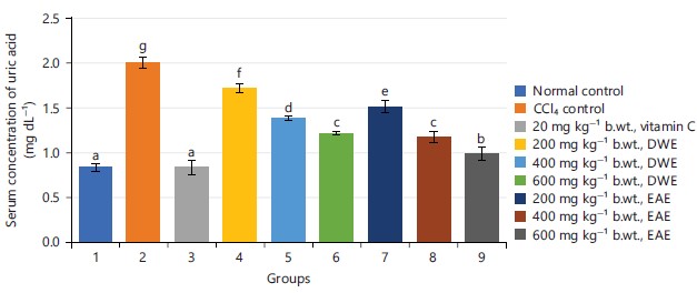
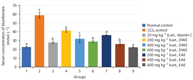
Effects of UCSE on serum renal function markers: The results of the effects of stem extracts of U. chamae (UCSE) on serum renal function markers of CCl4-induced toxicity in albino rats were presented in Fig. 1-6. The concentrations of serum urea, creatinine, uric acid, sodium, potassium and Bars with different letters are considered significantly (p<0.05) different, values are ±SEM, n = 5, UCSE: U. chamae stem extracts, DWE: Deionized water extract and EAE: Ethyl acetate extract bicarbonate were significantly (p<0.05) higher in the CCl4-administered untreated group compared to that of the normal control group.
|
|
|

|

|
|
Upon treatment with 200, 400 and 600 mg kg–1 b.wt., of UCSE, respectively, the concentrations of serum urea, creatinine, uric acid, sodium, potassium and bicarbonate were significantly (p<0.05) lower compared to the untreated group, with higher effects observed in the EAE than the DWE. The effects were found to be dose-dependent. Also, there were no significant (p<0.05) differences in the values of the group treated with 600 mg kg–1 b.wt., of EAE and the group treated with 20 mg kg–1 b.wt., of vitamin C.
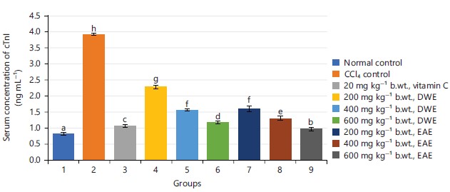
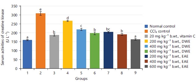
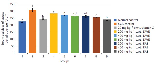
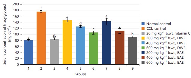
Effects of UCSE on serum cardiovascular function markers: Figure 7-13 show the results of the effects of UCSE on serum cardiovascular function markers of CCl4-induced toxicity in albino rats. There were significantly (p<0.05) higher activities of serum creatine kinase (CK) and lactate dehydrogenase, with significantly (p<0.05) higher concentrations of serum Cardiac Troponin I (cTnI), triacylglycerol (TG), cholesterol (TC) and Low-Density Lipoprotein Cholesterol (LDL), while the concentration of high-density lipoprotein cholesterol was significantly (p<0.05) lower in CCl4-administered untreated group compared to that of the normal control group. However, after treatment with different doses of UCSE for 21 days, it was found that the activities of serum creatine kinase (CK) and lactate dehydrogenase were significantly (p<0.05) lower, with significantly (p<0.05) lower concentrations of serum Cardiac Troponin I (cTnI), triacylglycerol (TG), cholesterol (TC) and Low-Density Lipoprotein Cholesterol (LDL), while the concentration of high-density lipoprotein cholesterol was significantly (p<0.05) higher when compared to the CCl4-administered untreated group. Higher effects were observed in the EAE than in the DWE. The effects were found to be dose-dependent. Moreover, there were no significant (p<0.05) differences in the values of the group treated with 600 mg kg–1 b.wt., of EAE and the group treated with 20 mg kg–1 b.wt., of vitamin C.
Effects of UCSE on the histology of the kidneys and hearts of the rats
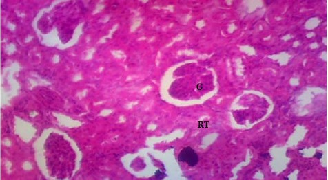
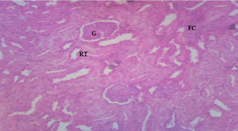
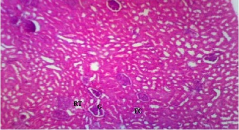
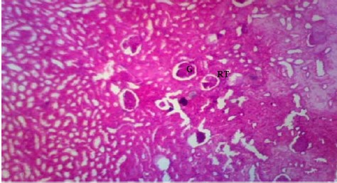
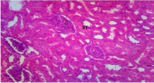
Kidney photomicrograph: The effects of the extracts on the kidney histology of the experimental rats were presented in Plate 1-9. The photomicrograph of the kidney section of group 1 rat (normal control) showed well-perfused and normal renal tissue architecture with normal tubular brush-borders (TB), normal glomeruli (G) in Bowman’s capsules (black arrow) surrounded by numerous renal tubules (RT) (Plate 1), whereas the kidney photomicrograph of group 2 administered with CCl4-without treatment (Plate 2) showed severe damage on the renal tissue (RT) with extensive fatty change (EFC), severe coagulative necrosis of glomeruli (G), loss of renal tissues in some areas and tubular necrosis (TN), infiltration of inflammatory cells (blue arrows) and loss of tubular brush-borders. However, mild regeneration of glomeruli (G) and renal tubules (RT) with mild fatty change (FC) was observed in groups 4, 5, 7 and 8 treated with 200 and 400 mg kg–1 b.wt., of DWE and EAE, respectively (Plate 4, 5, 7 and 8). While, the photomicrographs of kidney sections of groups 6 and 9, respectively treated with 600 mg kg–1 b.wt., of DWE and EAE (Plate 6 and 9) showed well-perfused renal tissue with moderate regeneration of glomeruli (G) and renal tubules (RT), together with group 3 treated with 20 mg kg–1 b.wt., of vitamin C (Plate 3).
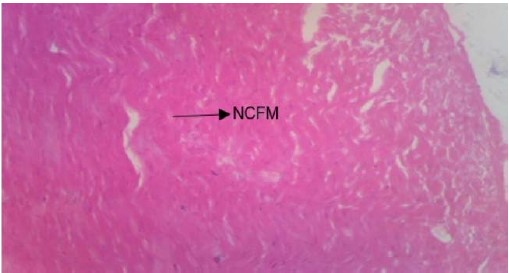
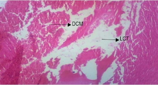
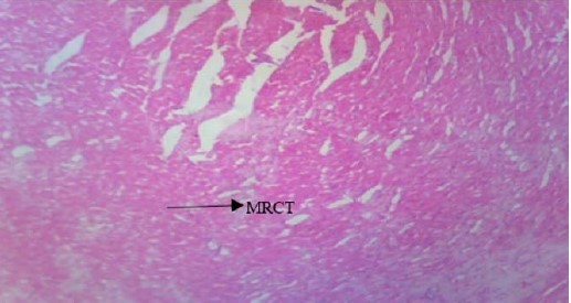
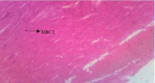
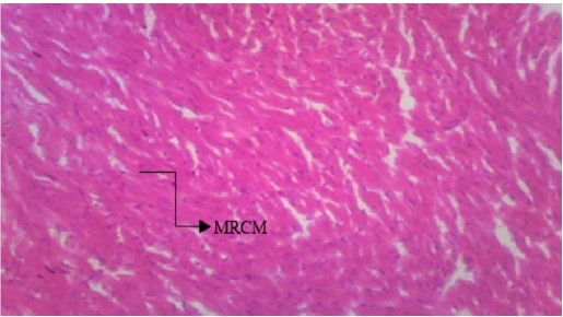
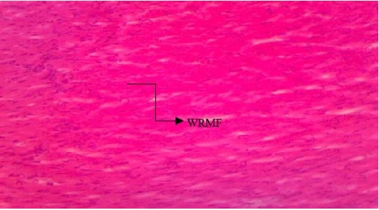
Heart photomicrograph: The effects of UCSE on the heart histology of the experimental rats were presented in Plate 10-18. The photomicrograph of the heart section of group 1 (normal control) showed normal cardiac fiber and muscle (NCFM) (Plate 10), whereas the heart photomicrograph of group 2 administered with CCl4-without treatment (Plate 11) showed disorganization of cardiac muscle (DCM) and loss of cardiac tissue (LCT). However, mild restoration of cardiac tissue (MRCT) was observed in groups 4 and 7 treated with 200 mg kg–1 b.wt., of DWE and EAE, respectively (Plate 13 and 16). The photomicrographs of heart sections of groups 5 and 6, respectively treated with 400 and 600 mg kg–1 b.wt., of DWE (Plate 14 and 15) showed moderate restoration of cardiac tissue (MRCT), as well as group 8 treated with 400 mg kg–1 b.wt., of EAE (Plate 17). Well-restored cardiac muscle and fibre (WRMF) were observed in groups 9 and 3 treated with 600 mg kg–1 b.wt., of EAE (Plate 18) and 20 mg kg–1 b.wt., of vitamin C (Plate 12) respectively.
|
|
|
|

|

|

|

|

|
|
|

|

|
|
|
|
|
|

|
|
|
|

|

|
|
DISCUSSION
The result showed that Carbon Tetrachloride (CCl4) induced nephrotoxicity and cardiotoxicity in the rats, indicated by alterations in the kidney and cardiovascular biomarkers. The results of the kidney function markers showed significant (p<0.05) increase in the concentrations of serum urea, creatinine, uric acid, Sodium (Na+), Potassium (K+) and Bicarbonate (HCO3–) in the CCl4-administered untreated group 2 compared to that of the normal control group 1, indicating that CCl4 might have induced kidney damage. Treatment of the CCl4-administered groups with 200, 400 and 600 mg kg–1 b.wt., of DWE and EAE of U. chamae stem respectively caused significant (p<0.05) reductions in the concentrations of the biomarkers in a dose-dependent manner (Fig. 1-6). Prevention of further progression of tissue damage and treatment of CCl4-induced kidney damage was also indicated by the kidney photomicrographs (Plate 1-9). Higher effects were observed in the EAE than DWE. Also, there were no significant (p<0.05) differences in the levels of urea, creatinine and K+ in group 9 treated with 600 mg kg–1 b.wt., of EAE and that of group 3 treated with 20 mg kg–1 b.wt., of vitamin C, while there were higher effects with significant (p<0.05) differences in the levels of Na+ and HCO3– in group 9 treated with 600 mg kg–1 b.wt., of EAE compared to group 3 treated with 20 mg kg–1 b.wt., of vitamin C. The results showed that CCl4 caused kidney damage in the albino rats, which was ameliorated to near-normal levels by U. chamae stem extracts. Hence, U. chamae stem possesses nephroprotective activity and could be suitable for the management of kidney problems. The findings were in agreement with previous reports on CCl4-induced kidney damage and its amelioration by plant extracts12,33-35.
Different studies have indicated that CCl4 intoxication causes free radical generation in many tissues including the liver, kidney, heart, lung, testis, brain and blood36-38. Therefore, processes involving reactive oxygen species (ROS) contribute significantly to the etiology of CCl4-induced damage in a vast array of biological systems and tissues, including the kidney36. In vitro and in vivo studies support the idea that CCl4 enhances lipid peroxidation and reduces the reduced/oxidized glutathione ratio in the kidney cortex as well as renal microsomes and mitochondria33. Serum urea and creatinine levels are the most commonly used screening tests for renal function. High serum levels of these metabolites indicate kidney problems. This is because the kidney is responsible for the filtration of the blood and when there is a problem with the kidney, urea and creatinine which are supposed to be filtered into the urine accumulate in the blood39,40. Moderate-to-advanced Chronic Kidney Disease (CKD) is commonly associated with elevated serum urea levels and studies have shown that urea is a direct and indirect uremic toxin, especially with regard to cardiovascular disease41. High blood levels of creatinine occur in urinary tract obstruction, chronic nephritis, etc., resulting in renal function impairment. Elevated levels of serum uric acid are observed in cases of hypertension and renal diseases42. The reduction in the levels of serum urea, creatinine, uric acid, Sodium (Na+), Potassium (K+) and bicarbonate in the CCL4-administered rats after treatment with extracts of U. chamae stem showed that the plant extracts, especially at 600 mg kg–1 b.wt., caused normal kidney function by increasing the renal reabsorption capacity of these substances. The results of the cardiovascular function markers showed significantly (p<0.05) higher activities of serum CK and LDH and concentrations of cTnI, TG, TC and LDL, with significant (p<0.05) reduction in HDL concentration in the CCl4-administered untreated group compared to that of the normal control group, which are indicators of Cardiovascular Diseases (CVDs). However, treatment of the CCl4-administered groups with different doses of UCSE resulted in significantly (p<0.05) lower levels of CK, LDH, cTnI, TG, TC, LDL and higher levels of HDL in a dose dependent manner, with the EAE-treated groups showing higher effects than their DWE group counterparts (Fig. 7-13). Thus, UCSE prevented further tissue damage in the rats administered with CCl4 and gradually reversed the toxic effects to near normal conditions, when compared to the untreated groups and the normal control, as shown in the heart photomicrographs (Plate 10-18). Therefore, current results showed that UCSE possesses cardioprotective activities and will be useful in the management of cardiovascular diseases. These observations were in agreement with previous studies on the effects of plant extracts on CCl4-induced cardiovascular diseases12,43,44.
The CCl4 is known to induce tissue damage through the generation of free radicals8. Tissue damages have been reported in fish exposed to herbicides45,46 and plant extracts47,48. Tissue damage leads to the leakage of its biomarkers from the cells into the bloodstream, leading to their serum increase, as evidenced by the increase in serum levels of cTnI, CK and LDH in the CCl4-administered rat groups. Cardiac Troponin (cTn) is one of the most commonly used biomarkers for myocardial injury. The troponin complex has 3 subunits-C, I and T that synergistically control calcium-mediated interaction of actin and myosin filament, leading to the contraction and relaxation of striated muscle or voluntary muscles49,50. About 3.5% of cTnI and 7% of cTnT exist freely in cardiac myocyte cytoplasm. The rest is bound within the sarcomere and is released as a result of proteolytic degradation. Myocardial Injury results in a disruption of the intracellular contractile proteins leading to an increase in the cytoplasmic pool content of cTnI and cTnT. Calcium and troponin control the production of cardiac muscular force. Increased cardiac muscle calcium concentration leads to muscle contraction27. Calcium is bound to troponin-C triggering activation of tropomyosin which in turn binds troponin-T to form troponin-tropomyosin complex. Cardiac troponin I now bind actin to link and sustain the troponin-tropomyosin complex in place, thereby maintaining normal cardiac muscle contraction and normal heart physiology. Troponin-I is cardiac specific27, while Troponin-T is derived from both the myocardium and skeletal muscles. The release of cardiac troponin I into the circulation is a manifestation of reversible or irreversible myocyte injury51.
Creatine kinase (CK) or Creatine Phosphokinase (CPK) mediates in the formation of creatine phosphate from creatine and ATP. There are three isoenzymes of CK with two subunits M (muscles) and B (brain) existing as CPK-BB, CPK-MB and CPK-MM. High plasma activity of CK indicates damage to skeletal muscle, which does not indicate acute myocardial infarction (AMI)50,52. However, about 40% of the CK activity is comprised of CPK-MB (or CK-2) in cardiac muscle and about 2% in most muscle groups and other tissues, being the first enzyme released into circulation 6-8 hrs after the infarction, reaching the peak value within 24-30 hrs and returns to normal level within 2-3 days, serving as a more specific and sensitive marker for MI53. Lactate Dehydrogenase (LDH) catalyzes the interconversion of lactate and pyruvate by NAD+-dependent reaction. It is tetrameric with two subunits: M (muscles) and H (Heart) and five isoenzymes LDH1, LDH2, LDH3, LDH4 and LDH5. The peak activities of LDH occur within 3 to 4days and remain elevated for up to 10days after myocardial infarction, indicating that the removal of extra serum LDH in blood circulation occurs slowly54. Also, altered lipid profile, as observed in our study such as elevated levels of TG, TC and LDL with low levels of HDL, is a sign of tissue damage such as cardiotoxicity.
Dyslipidemia is the most common cause of cardiovascular diseases. It leads to the deposition of lipid on the arterial wall, thereby aggravating the process of atherosclerosis55 and is associated with high TC, TG, LDL, with low HDL levels in plasma56,57. Several studies have reported increased levels of serum TC, TG and LDL, with decreased levels of HDL in coronary heart disease58,59 and diabetic patients60. The medicinal activities of U. chamae have been attributed to its various phytochemical constituents15. Also, a poly-herbal drug, Ruzu herbal bitters (RHB), containing Uvaria chamae, was shown to possess several bioactive compounds with antihyperlipidemic, antidiabetic, antioxidant, antibiotic, anticancer, hepatoprotective and nephroprotective properties61.
This study happened to be the first study on the effects of deionized water and ethyl acetate extracts of Uvaria chamae stem on renal and cardiovascular parameters of Carbon Tetrachloride (CCl4)-induced toxicity in rats. The study was necessary since renal and cardiovascular diseases are among the leading causes of death, with a high cost of treatment using orthodox drugs which have several side effects. Therefore, the findings bring to the public knowledge that U. chamae stem extracts possess nephroprotective and cardioprotective activities and could be harnessed for the management of renal and cardiovascular diseases, being cheaper and locally available. The study also has limitations, as it has to be carried out in humans to ascertain similar effects in renal and cardiac patients compared to CCl4 induction in rats, including the doses. Hence, this study could serve as a hypothesis that has to be proven further to form a theory. It is therefore recommended that further study into the very bioactive substances responsible for these activities in the plant be carried out and harnessed into medication.
CONCLUSION
Carbon Tetrachloride (CCl4)-administration in rats resulted in alterations in renal and cardiovascular parameters such as serum levels of urea, creatinine, uric acid, Sodium (Na+), Potassium (K+) and Bicarbonate (HCO3–), creatine kinase (CK), Lactate Dehydrogenase (LDH), Cardiac Troponin I (cTnI), triacylglycerol (TG), total cholesterol (TC), Low-Density Lipoprotein Cholesterol (LDL) and High-Density Lipoprotein Cholesterol (HDL), indicating that CCl4 might have induced renal and cardiovascular damages. These effects were prevented and gradually reversed to near-normal levels after treatment of CCl4-administered rat groups with different doses of deionized water and ethylacetate stem extracts of Uvaria chamae. The ethyl acetate extract showed higher efficacy than the deionized water extract. The result of this study showed that Uvaria chamae stem possesses nephroprotective and cardioprotective activities. Therefore, the bioactive compounds could be pharmacologically useful in the management and prevention of renal and cardiovascular diseases.
SIGNIFICANCE STATEMENT
Renal and cardiovascular diseases are among the leading causes of death worldwide. Because of the high cost and side effects of orthodox drugs, there is need for a less costly and more effective herbal medicines with antioxidant and anti-inflammatory activities for the treatment of these diseases. This study investigated the effects of deionized water and ethyl acetate extracts of Uvaria chamae stem on renal and cardiovascular parameters of Carbon Tetrachloride (CCl4)-induced toxicity in albino rats. The ethyl acetate extract showed higher efficacy than the deionized water extract. The findings of this study showed that the Uvaria chamae stems possess nephroprotective and cardioprotective activities. Thus, the bioactive compounds could be pharmacologically useful in the management and prevention of renal and cardiovascular diseases.
REFERENCES
- Perazella, M.A., 2009. Renal vulnerability to drug toxicity. Clin. J. Am. Soc. Nephrol., 4: 1275-1283.
- Hensle, T.W. and E.H. Lambert, 2010. Renal Function, Fluids, Electrolytes, and Nutrition from Birth to Adulthood. In: Pediatric Urology, Gearhart, J.P., R.C. Rink and P.D.E. Mouriquand (Eds.), W.B. Saunders, Philadelphia, ISBN: 9781416032045, pp: 23-30.
- Radi, Z.A., 2019. Kidney pathophysiology, toxicology, and drug-induced injury in drug development. Int. J. Toxicol., 38: 215-227.
- Barnett, L.M.A. and B.S. Cummings, 2018. Nephrotoxicity and renal pathophysiology: A contemporary perspective. Toxicol. Sci., 164: 379-390.
- Celermajer, D.S., C.K. Chow, E. Marijon, N.M. Anstey and K.S. Woo, 2012. Cardiovascular disease in the developing world: Prevalences, patterns and the potential of early disease detection. J. Am. Coll. Cardiol., 60: 1207-1216.
- Khan, I.A., M. Hussain, S.K. Syed, M. Saadullah and A.M. Alqahtani et al., 2022. Pharmacological justification for the medicinal use of Plumeria rubra Linn. in cardiovascular disorders. Molecules, 27.
- Khan, I.A., M. Hussain, S.H. Munawar, M.O. Iqbal and S. Arshad et al., 2021. Jasminum sambac: A potential candidate for drug development to cure cardiovascular ailments. Molecules, 26.
- Adaramoye, O.A., 2009. Comparative effects of vitamin E and kolaviron (a biflavonoid from Garcinia kola) on carbon tetrachloride-induced renal oxidative damage in mice. Pak. J. Biol. Sci., 12: 1146-1151.
- Shenoy, K.A., S.N. Somayaji and K.L. Bairy, 2001. Hepatoprotective effects of ginkgo biloba against carbon tetrachloride induced hepatic injury in rats. Indian J. Phamacol., 33: 260-266.
- Adewole, S.O., A.A. Salako, O.W. Doherty and T. Naicker, 2007. Effect of melatonin on carbon tetrachloride-induced kidney injury in Wistar rats. Afr. J. Biomed. Res., 10: 153-164.
- Plaa, G.L. and H. Witschi, 1976. Chemicals, drugs and lipid peroxidation. Annu. Rev. Pharmacol. Toxicol., 16: 125-142.
- Agbafor, K.N., C. Ezeali, E.I. Akubugwo, I.K. Obiudu and A.J. Uraku et al., 2015. Cardioprotective effect of leaf and root extracts of Newbouldia laevis against carbon tetrachloride induced-cardiotoxicity in albino rats. Eur. J. Med. Plants, 9.
- Giordano, F.J., 2005. Oxygen, oxidative stress, hypoxia, and heart failure. J. Clin. Invest., 115: 500-508.
- Irvine, F.R., 1961. Woody Plants of Ghana: With Special Reference to Their Uses. Oxford University Press, Oxford, England, Pages: 868.
- Obasi, D.C. and J.N. Obasi, 2021. Comparative study on the levels of phytochemicals present in n-hexane, ethanol, aqueous, ethylacetate and methanol extracts of Uvaria chamae stem. J. Med. Stud., 9: 195-200.
- Oliver-Bever, B., 1986. Medicinal Plants in Tropical West Africa. Cambridge University Press, Cambridge, ISBN: 9780521268158, Pages: 375.
- Igoli, J.O., O.G. Ogaji, T.A. Tor-Anyiin and N.P. Igoli, 2005. Traditional medicine practice amongst the Igede people of Nigeria. Part II. Afr. J. Tradit. Complimentary Altern. Med., 2: 134-152.
- Thomas, P.S. and E.E. Essien, 2020. Antiglycation, antioxidant, and cytotoxic activities of Uvaria chamae root and essential oil composition. Nat. Prod. Res., 34: 880-883.
- Okwu, D.E. and F. Iroabuchi, 2009. Phytochemical composition and biological activities of Uvaria chamae and Clerodendoron splendens. J. Chem., 6: 553-560.
- James, O., E.U. Godwin and I.G. Otini, 2013. Uvaria chamae (Annonaceae) plant extract neutralizes some biological effects of Naja nigricollis snake venom in rats. Br. J. Pharmacol. Toxicol., 4: 41-50.
- Quinones, M., M. Miguel and A. Aleixandre, 2013. Beneficial effects of polyphenols on cardiovascular disease. Pharmacol. Res., 68: 125-131.
- Lorke, D., 1983. A new approach to practical acute toxicity testing. Arch. Toxicol., 54: 275-287.
- Tietz, N.W., 2006. Tietz Textbook of Clinical Chemistry and Molecular Diagnostic. 4th Edn., Elsevier Saunders, New York, ISBN: 9780721601892, Pages: 2412.
- Fossati, P., L. Prencipe and G. Berti, 1980. Use of 3,5-dichloro-2-hydroxybenzenesulfonic acid/4-aminophenazone chromogenic system in direct enzymic assay of uric acid in serum and urine. Clin. Chem., 26: 227-231.
- Tietz, N.W., 1976. Fundamentals of Clinical Chemistry, 2nd Edn., Saunders, London, United States, ISBN: 9780721688664, Pages: 1263.
- Forrester, R.L., L.J. Wataji, D.A. Silverman and K.J. Pierre, 1976. Enzymatic method for determination of CO2 in serum. Clin. Chem., 22: 243-245.
- Adams, J.E., G.S. Bodor, V.G. Davila-Roman and J.A. Delmez, 1993. Cardiac troponin I. A marker with high specificity for cardiac injury. Circulation, 88: 101-106.
- Szasz, G., W. Gruber and E. Bernt, 1976. Creatine kinase in serum: 1. Determination of optimum reaction conditions. Clin. Chem., 22: 650-656.
- Friedman, R.B. and D.S. Young, 1997. Effects of Disease on Clinical Laboratory Tests. 3rd Edn., American Association for Clinical Chemistry Press, Washington, DC, ISBN: 9780915274871, Pages: 1068.
- Expert Panel on Detection, Evaluation and Treatment of High Blood Cholesterol in Adults, 2001. Executive summary of the third report of the National Cholesterol Education Program (NCEP) expert panel on detection, evaluation and treatment of high blood cholesterol in adults (Adult Treatment Panel III). J. Am. Med. Assoc., 285: 2486-2497.
- Tietz, N.W., 1995. Clinical Guide to Laboratory Tests. 3rd Edn., W.B. Saunders Company, Philadelphia, USA, ISBN: 072165035X, Pages: 1096.
- Friedewald, W.T., R.I. Levy and D.S. Fredrickson, 1972. Estimation of the concentration of low-density lipoprotein cholesterol in plasma, without use of the preparative ultracentrifuge. Clin. Chem., 18: 499-502.
- Yilmaz-Ozden, T., A. Can, A. Karatug, Z. Pala-Kara, A. Okyar and S. Bolkent, 2014. Carbon tetrachloride-induced kidney damage and protective effect of Amaranthus lividus L. in rats. Toxicol. Ind. Health, 32: 1143-1152.
- Awodele, O., A.A. Adeneye, S.A. Aiyeola and A.S. Benebo, 2015. Modulatory effect of Mangifera indica against carbon tetrachloride induced kidney damage in rats. Interdiscip. Toxicol., 8: 175-183.
- Iseghohi, S.O. and N.E.J. Orhue, 2017. Aqueous extract of Dennettia tripetala ameliorates liver and kidney damage caused by multiple exposures to carbon tetrachloride. Clin. Phytosci.
- Kumar, G., G.S. Banu and M.R. Pandian, 2005. Evaluation of the antioxidant activity of Trianthema portulacastrum L. Indian J. Pharmacol., 37: 331-333.
- Khan, M.R. and D. Ahmed, 2009. Protective effects of Digera muricata (L.) Mart. on testis against oxidative stress of carbon tetrachloride in rat. Food Chem. Toxicol., 47: 1393-1399.
- Khan, M.R., W. Rizvi, G.N. Khan, R.A. Khan and S. Shaheen, 2009. Carbon tetrachloride-induced nephrotoxicity in rats: Protective role of Digera muricata. J. Ethnopharmacol., 122: 91-99.
- Al-Yahya, M., R. Mothana, M. Al-Said, M. Al-Dosari and N. Al-Musayeib et al., 2013. Attenuation of CCl4-induced oxidative stress and hepatonephrotoxicity by Saudi Sidr honey in rats. Evidence-Based Complementary Altern. Med., 2013.
- Mahmoud, A.M., R.R. Ahmed, H.A. Soliman and M. Salah, 2015. Ruta graveolens and its active constituent rutin protect against diethylnitrosamine-induced nephrotoxicity through modulation of oxidative stress. J. Appl. Pharm. Sci., 5: 16-21.
- Laville, S.M., A. Couturier, O. Lambert, M. Metzger and N. Mansencal et al., 2023. Urea levels and cardiovascular disease in patients with chronic kidney disease. Nephrol. Dialysis Transplant., 38: 184-192.
- Kanbay, M., Y. Solak, E. Dogan, M.A. Lanaspa and A. Covic, 2010. Uric acid in hypertension and renal disease: The chicken or the egg? Blood Purif., 30: 288-295.
- Nwidu, L.L., Y.I. Oboma, E. Elmorsy and W.G. Carter, 2018. Alleviation of carbon tetrachloride-induced hepatocellular damage and oxidative stress with a leaf extract of Glyphae brevis (Tiliaceae). J. Basic Clin. Physiol. Pharmacol., 29: 609-619.
- Ude, C.M. and K.N. Agbafor, 2018. Comparative study on the cardio protective properties of leaf and seed extracts of Persea americana against carbon tetrachloride induced cardiotoxicity in albino rats. Int. Digital Organ. Sci. Res., 3: 88-101.
- Ikechukwu, A.A., U.A. Ibiam, U.O. Obasi, D.O. Chukwu, P.C.U. Okechukwu and O.R. Inya-Agha, 2015. Evaluation of the antioxidant properties of aqueous stem bark extract of the Bridelia ferruginea and butachlor. Global J. Pharmacol., 9: 352-357.
- Obasi, D.C., U.A. Ibiam, J.N. Obasi and I.A. Ali, 2021. Biochemical effects of glyphosate on juvenile African catfish (Clarias gariepinus). Int. J. Sci. Res. Eng. Dev., 4: 1472-1482.
- Ama, I.U., C.O. David, O.U. Orji, A.P. Maduabuchi, C. Chukwu and J.N. Obasi, 2015. GC-MS analysis, acute toxicity and oxidative stress potentials (effects) of Albizia chevalieri extract on juvenile African catfish (Clarias gariepinus). Middle-East J. Sci. Res., 23: 192-199.
- Obasi, D.C., J.N. Obasi, I.A. Ali and U.A. Ibiam, 2022. Toxicity of aqueous stem-bark extract of Albizia chevalieri on the liver and kidney of juvenile African catfish (Clarias gariepinus). J. Biol. Sci., 22: 42-49.
- Daubert, M.A. and A. Jeremias, 2010. The utility of troponin measurement to detect myocardial infarction: Review of the current findings. Vasc. Health Risk Manage., 6: 691-699.
- Aydin, S., K. Ugur, S. Aydin, İ. Sahin and M. Yardim, 2019. Biomarkers in acute myocardial infarction: Current perspectives. Vasc. Health Risk Manage., 15: 1-10.
- Reichlin, T., W. Hochholzer, S. Bassetti, S. Steuer and C. Stelzig et al., 2009. Early diagnosis of myocardial infarction with sensitive cardiac troponin assays. N. Engl. J. Med., 361: 858-867.
- van der Veen, K.J. and A.F. Willebrands, 1966. Isoenzymes of creatine phosphokinase in tissue extracts and in normal and pathological sera. Clin. Chim. Acta, 13: 312-316.
- Roberts, R., K.S. Gowda, P.A. Ludbrook and B.E. Sobel, 1975. Specificity of elevated serum MB creatine phosphokinase activity in the diagnosis of acute myocardial infarction. Am. J. Cardiol., 36: 433-437.
- Wu, Y., C. Lu, N. Pan, M. Zhang and Y. An et al., 2021. Serum lactate dehydrogenase activities as systems biomarkers for 48 types of human diseases. Sci. Rep., 11.
- Garg, R., S. Aggarwal, R. Kumar and G. Sharma, 2015. Association of atherosclerosis with dyslipidemia and co-morbid conditions: A descriptive study. J. Nat. Sci. Biol. Med., 6: 163-168.
- Sherbet, D.P., P. Garg, E.S. Brilakis and S. Banerjee, 2013. Low-density lipoprotein cholesterol: How low can we go?. Am. J. Cardiovasc. Drugs, 13: 225-232.
- Kullisaar, T., K. Zilmer, T. Salum, A. Rehema and M. Zilmer, 2016. The use of probiotic L. fermentum ME-3 containing Reg’Activ Cholesterol supplement for 4 weeks has a positive influence on blood lipoprotein profiles and inflammatory cytokines: An open-label preliminary study. Nutr. J., 15.
- Shabana, S.U. Shahid and S. Sarwar, 2020. The abnormal lipid profile in obesity and coronary heart disease (CHD) in Pakistani subjects. Lipids Health Dis., 19.
- Dabas, A., S. Yadav and V.K. Gupta, 2014. Lipid profile and correlation to cardiac risk factors and cardiovascular function in type 1 adolescent diabetics from a developing country. Int. J. Pediatr., 2014.
- Obasi, D.C. and V.N. Ogugua, 2021. Effect of ruzu herbal bitters on the liver function and lipid profile parameters of alloxan-induced diabetic rats. J. Clin. Exp. Hepatol.
- Obasi, D.C. and V.N. Ogugua, 2021. GC-MS analysis, pH and antioxidant effect of ruzu herbal bitters on alloxan-induced diabetic rats. Biochem. Biophys. Rep., 27.
How to Cite this paper?
APA-7 Style
Obas,
J.N., Obasi,
D.C., Agbafo,
K.N. (2023). Uvaria chamae Stem Extracts Possess Nephroprotective and Cardioprotective Effects Against Carbon Tetrachloride-Induced Toxicity in Albino Rats. Asian Journal of Biological Sciences, 16(4), 692-712. https://doi.org/10.3923/ajbs.2023.692.712
ACS Style
Obas,
J.N.; Obasi,
D.C.; Agbafo,
K.N. Uvaria chamae Stem Extracts Possess Nephroprotective and Cardioprotective Effects Against Carbon Tetrachloride-Induced Toxicity in Albino Rats. Asian J. Biol. Sci 2023, 16, 692-712. https://doi.org/10.3923/ajbs.2023.692.712
AMA Style
Obas
JN, Obasi
DC, Agbafo
KN. Uvaria chamae Stem Extracts Possess Nephroprotective and Cardioprotective Effects Against Carbon Tetrachloride-Induced Toxicity in Albino Rats. Asian Journal of Biological Sciences. 2023; 16(4): 692-712. https://doi.org/10.3923/ajbs.2023.692.712
Chicago/Turabian Style
Obas, Jennifer, Nkeiru, David Chukwu Obasi, and Kingsley Nwonu Agbafo.
2023. "Uvaria chamae Stem Extracts Possess Nephroprotective and Cardioprotective Effects Against Carbon Tetrachloride-Induced Toxicity in Albino Rats" Asian Journal of Biological Sciences 16, no. 4: 692-712. https://doi.org/10.3923/ajbs.2023.692.712

This work is licensed under a Creative Commons Attribution 4.0 International License.