Evaluation of the Effect of an Aqueous Extract of Psidium guajava (Guava) Leaves on the Frontal Cortex of Diabetic Wistar Rats
-
Isabel Namfukwe Luambia
Department of Human Anatomy, School of Medicine and Health Sciences, Mulungushi University, Livingstone Campus, Zambia
Ally SiabwachaDepartment of Human Anatomy, School of Medicine and Health Sciences, Mulungushi University, Livingstone Campus, Zambia
Memory NgosaPharmacy Unit, School of Medicine and Health Sciences, Mulungushi University, Livingstone Campus, Zambia
Sharon kaunduDepartment of Clinical sciences, School of Medicine, Eden University, Lusaka, Zambia
Mwitwa kombeDepartment of Clinical sciences, School of Medicine, Eden University, Lusaka, Zambia
Lukundo Mulambia SiameDepartment of Human Anatomy, School of Medicine and Health Sciences, Mulungushi University, Livingstone Campus, Zambia
Mulenga MalataZambia Compulsory Standards agency, Lusaka, Zambia
Uthman Ademola Yusuf
Department of Human Anatomy, School of Medicine and Health Sciences, Mulungushi University, Livingstone Campus, Zambia
| Received 22 Feb, 2024 |
Accepted 04 Apr, 2024 |
Published 30 Jun, 2024 |
Background and Objective: Psidium guajava is a medicinal plant used for the treatment and management of numerous ailments including diabetes mellitus. This study aimed to investigate the antihyperglycemic effect of aqueous extract of Psidium guajava on the frontal cortex of diabetic Wistar rats. Materials and Methods: Thirty male adult Wistar rats were randomly selected into five groups made up of six Wistar rats each, control, diabetic control, diabetic+metformin, diabetic+guava and guava only groups. The induction of diabetes was done using streptozotocin at a dose of 70 mg/kg b.wt. and undergo elevation in blood glucose levels, after 72 hrs of established hyperglyemia treatment begin with aqueous guava extract at 400 mg/kg b.wt. and metformin at 100 mg/kg b.wt., respectively and was given orally for 4 weeks. Results: The average body weight and relative brain weight of the diabetic+guava and diabetic+metformin was significant when compared to the diabetic group (p<0.05). Diabetic+guava became normoglycemic at week 3 while diabetic+metformin at week 4. The histological examination of frontal cortex of the diabetic rats showed disruption in histoarchitecture with necrotic astrocyte and pyramidal cells. Diabetic+guava and diabetic+metformin revealed little damage in the astrocyte and pyramidal cells in their histoarchitecture. Guava only was similar to control. The analysis of the reduced Glutathione (GSH), Glucose-6-Phosphate Dehydrogenase (G-6-PDH) and Lactate Dehydrogenase (LDH) activities in the frontal cortex revealed that the rats in the diabetic+guava leaves and diabetic+metformin are similar to the control group. Conclusion: The aqueous extract of guava leaves showed remedial effects on the frontal cortex of adult male Wistar rats against the destruction caused by diabetes.
| Copyright © 2024 Luambia et al. This is an open-access article distributed under the Creative Commons Attribution License, which permits unrestricted use, distribution, and reproduction in any medium, provided the original work is properly cited. |
INTRODUCTION
Diabetes mellitus is a chronic disorder that follows when the pancreas produces inadequate insulin or when the body cannot efficiently utilize the insulin it produces1. According to the IDF diabetes atlas of 2021, it was reported that almost one in two adults (20-79 years old) had undiagnosed diabetes (44.7%; 239.7 million). Western Pacific reported (52.8%) and South-East Asia regions (51.3%), respectively. The least percentage of undiagnosed diabetes was detected in North America and the Caribbean (24.2%). The highest proportions of these cases (53.6%) were found in Africa2.
In Sub-Saharan Africa (SSA) complications accounted for chronic Noncommunicable Diseases (NCDs) such as diabetes mellitus are on a steady increase, it was approximated that, in 2014, 20 million people in SSA were diabetic and 523,000 deaths occurred because of this disease or conditions related to it, with 76% of them under the age of 603.
It was predicted that increased age and urbanization of the population favor a continued upward trend of diabetes mellitus in the region with a prediction of 41.5 million people becoming diabetic by the year 2035, in SSA. This also poses a substantial socioeconomic problem in the face of limited resources for government and the affected population. In relation to prevalence, the inadequate data available reveals that the prevalence varies from 3 to 15% as an overall diabetes prevalence for SSA in 20134.
Left untreated diabetes mellitus can lead to serious complications among them neuropathies and clinical syndromes such as ketosis and can also be linked with Alzheimer’s Disease (AD) which is a neurodegenerative disorder5.
There is a vast use of traditional medicine in Africa as reported by the WHO Global Report on Traditional and Complementary Medicine of 2019 which indicated an 85% of the total member states in the WHO African Region confirming the use of traditional medicine in the treatment of different diseases including diabetes mellitus. Psidium guajava traditionally has been used in the treatment of diabetes mellitus due to its availability in Africa6. It is under this pretext that this research sought to investigate the antidiabetic effects of guava leaf extract which has been used in folk medicine for the treatment of different disorders including diabetes mellitus.
MATERIALS AND METHODS
Study area: The study was carried out at the Department of Human Anatomy Mulungushi University School of Medicine and Health Sciences, Livingstone Campus, Zambia between April and September, 2023.
Plant materials: The guava leaves were collected from Kabwe district, Central Province of Zambia. It was subjected to identification at the University of Zambia School of Natural Sciences under the Department of Biological Sciences before the study begun. The guava leaves were air-dried and pounded. The dry pounded guava leaves were then grounded and sieved to obtain a homogenous powder (800 g). The extraction was done using Soxhlet extraction methods7.
Animals and animal management: Thirty adult presumably healthy male Wistar rats (Rattus norvegicus) were used for this study. The animals were between 8 to 10 weeks old; body weight (160-200 g). Animals were kept in five cages (6 rats per cage) and housed in the Animal House of, Mulungushi University School of Medicine and Health Sciences, Nursing Department, Kabwe town campus. They were maintained on standard animal feeds (Wealth-gate pelletized feeds) and were given access to clean water and feeds freely.
Induction of diabetes: Diabetes mellitus was induced using Streptozotocin (STZ). Rats were weighed and a baseline glucose level was initiated after the overnight fasting period. The animals were inoculated with STZ calculated at a dose of 70 mg/kg b.wt. and reintroduced to the normal feeding cycle7. It takes about 72 hrs for diabetes to be established in the animals following the administration of STZ therefore a fasting blood sugar was collected to determine the onset of diabetes by means of a tail vein puncture 72 hrs following administration of STZ. Accu-Chek glucometer (Mannheim, Germany) was used to estimate blood glucose levels. The rats were considered diabetic with fasting blood glucose levels above 7 mmol/L/ >250 mg/dL.
Experimental design: Thirty adult male Wistar rats were randomly selected into five groups of six. Control Animals in group A were normoglycemic and got neither STZ nor guava extract; those in group B were normal and received guava extract only; those in group C were treated for diabetes with guava extract; and those in group D were treated for diabetes with metformin and those in group E were diabetic but received neither guava nor metformin.
Guava mode of administration: The physiological saline was used to dissolve guava leaves on a daily basis and administered orally using an orogastric cannula to group B and C rats at 400 mg/kg b.wt., for 4 weeks, group D received metformin at 100 mg/kg b.wt. Group A and E rats received neither STZ nor guava leaf extract8.
Measurement of blood glucose level: Glucose oxidase method of one touch ultra 2 glucometer (Accu-Chek Compact Plus) was used to evaluate blood glucose in overnight fasted Wistar rats between 9:00-10:00 hrs. Blood was obtained from the median caudal vein of the tail by snipping the tip of the tail. The blood glucose level was monitored weekly for 2 weeks (acclimatisation period) before the induction of diabetes and for four weeks of treatment8.
Measurement of the body weight (g): Body weight (g) of the rats was recorded for two weeks (acclimatisation period) prior to induction of diabetes and on a weekly basis during the experimental treatment for a period of four weeks. Body Weight was taken with a weighing scale (Venus VT 30 SL)8.
Relative organ weight (%): The relative organ weight of the rat was evaluated as the ratio of respective weight of the brain and the terminal body weight of the same rat, the unit was recorded as percentage using a sensitive weighing balance (SonyF3G brand)8.
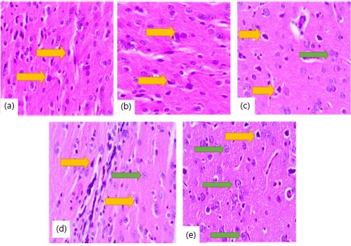
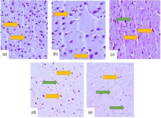
Histological and histochemical studies: At the end of this study, animals were sacrificed by euthanasia. They were laid in a supine position on the dissecting board and pinned through the fore and hind paws. The skulls of the animals were dissected with bone forceps and each organ was carefully removed and weighed. The tissue for histological studies was fixed in freshly prepared formal saline for 72 hrs and processed for routine histological examinations stained with Hematoxylin and Eosin (H&E) to observe changes in the cellular morphology and PTAH for observing the astrocyte. The tissues for enzymes of glucose metabolism (G6PDH and LDH) and oxidative stress markers (GSH) studies were immediately placed in 0.1M of phosphate buffer solution (PH 7.4) for homogenization9.
Photomicrography: Photomicrography of histological sections of the prefrontal cortices was taken with an Olympus Microscope (New York, United States of America) coupled with a camera at Department of Human Anatomy, Mulungushi University School of Medicine and Health Sciences, Livingstone Campus, Zambia.
Statistical analysis: Data was presented as Mean±Standard error of the mean (Mean±SEM); analysed using one-way ANOVA. All graphs were drawn using Excel (Microsoft Corporation, USA). The p-values less than 0.05 (p<0.05) were taken to be statistically significant.
Ethical consideration: Ethical and regulatory approval was obtained from Mulungushi University School of Medicine and Health Sciences Research Ethics Committee and the National Health Research Authority, Zambia.
RESULTS
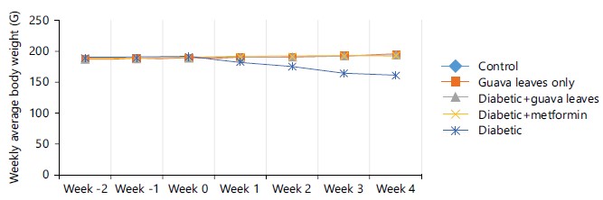
Figure 1 shows weekly changes in average body weight of Wistar rats belonging to different groups. In the weeks of acclimatization (week -1 and -2), the body weight was similar in all the groups when compared to each other there was no significant (p>0.05). After induction (week 0), the body weight of Wistar rats in the diabetic group was maintained without significant change when compared to the control group. In week 3 there was a significant decrease in the diabetic group rats when compared to the control, diabetic+guava leaves, diabetic+metformin and guava leaves only groups which was significant (p<0.05). In week 4 diabetic group rats continued showing a decline in body weight when compared to the control, guava leaves only, diabetic+guava leaves, diabetic+metformin it was significant (p<0.05).
Figure 2 shows the relative weight of the brain in different groups of Wistar rats. The diabetic group showed a significant decline in the relative brain weight as compared to other groups (p<0.05). The rats in the diabetic+guava leaves and diabetic+metformin groups show a slight decline as compared to the control and guava leaves only groups but was not significant (p>0.05).
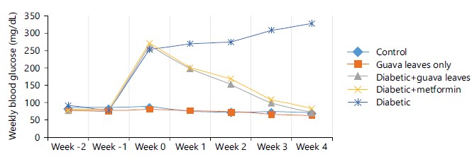
Figure 3 displays blood glucose levels which were recorded weekly in various groups of Wistar rats. During the acclimatization weeks the blood glucose levels of all groups were normal without significant difference when compared to control group (p>0.05). There was an upsurge in blood glucose during the week of induction (week 0) in diabetic, diabetic+guava leaves and diabetic+metformin groups. Following treatment in week 1 diabetic+guava leaves and diabetic+metformin revealed a significant decline when compared to diabetic group which was significant (p<0.05). In the week 3 of treatment diabetic+guava leaves had achieved normal glycemic level and it was significant when compared to diabetic group (p<0.05). In week 4 both diabetic+guava leaves and diabetic+metformin groups were significant to diabetic group (p<0.05); when diabetic+guava leaves and diabetic+metformin groups are compared to control group there was no significant (p>0.05).
|

|
|
|
Histology of the frontal cortex
Hematoxylin and Eosin (H&E) stain: In the control and guava-only groups, the pyramidal cells appear normal. The cells are densely populated and the histoarchitecture of the frontal cortices was preserved (Fig. 4a-b). Diabetic+guava and diabetic+metformin groups, some cells were necrotic while some cells appeared normal (Fig. 4c-d). In the diabetic group, most of the cells present were necrotic and the normal cells present were few (Fig. 4e).
Phosphotungstic acid hematoxylin stain: The rats in the control and guava-only groups showed normal distribution of astrocytes and the histoarchitecture appeared normal (Fig. 5a-b). Diabetic+guava, diabetic+metformin and diabetic groups, some neurons were degenerated while some appeared normal with astrocytes (Fig. 5c-e).
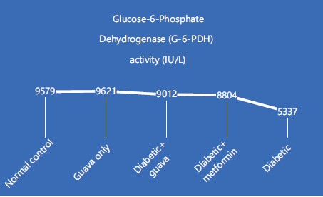
Glucose-6-Phosphate Dehydrogenase (G-6-PDH) activity in the frontal cortex (IU/L): The activity of Glucose-6-Phosphate Dehydrogenase (G-6-PDH) in various groups of Wistar rats illustrated in Fig. 6 shows the rats in the guava only group had the highest G-6-PDH activity while the rats in the diabetic group had the smallest activity. When the control and guava-only group was compared to the diabetic group the decrease was significant (p<0.05). Both the diabetic+guava leaves and the diabetic+metformin groups had similar G-6-PDH activity when compared to the control and guava only groups there was no significant record (p>0.05).
|
|
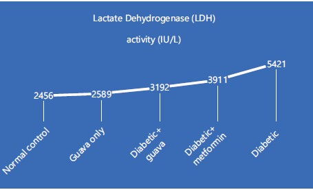
Lactate Dehydrogenase (LDH) activity in frontal cortex (IU/L): Figure 7 shows the activity of Lactate Dehydrogenase (LDH) in different groups of Wistar rats. The diabetic group showed a significant upsurge in the activity of lactate dehydrogenase as compared to control and guava-only groups, it was significant when compared (p<0.05). The rats in the diabetic+guava leaves and diabetic+metformin groups show a slight increase in contrast to the control and guava leaves only groups when compared it was not significant (p>0.05).
|
|
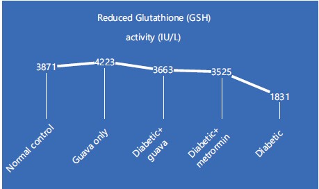
Reduced Glutathione (GSH) activity in the frontal cortex (IU/L): Figure 8 displays the reduced Glutathione (GSH) activity in various groups of Wistar rats. The rats in the diabetic group had the least GSH activity while guava only group had the highest. The diabetic+guava leaves and diabetic+metformin groups are in the same range of activity. When the rats in diabetic group were compared to the control, guava-only, diabetic+guava leaves and diabetic+metformin groups there was a significant decrease (p<0.05). When diabetic+guava leaves and diabetic+metformin groups were compared to control and guava groups the decrease was not significant (p>0.05).
DISCUSSION
Guava leaves aqueous extract has been reported as having antihyperglycemic and antidiabetic effects, hence its use in the traditional management of hyperglycemia10.
In this present study, the average body weight of diabetic group rats shows that there was a decrease from week 1 to week 4 when compared to other groups it was significant this might be due to gluconeogenesis11 while the diabetic+guava leaves and diabetic+metformin groups are maintained the average bodyweight when compared to the control group of rats it was not significant this is due to the suppression of the gluconeogenesis by the guava leaves extract and the metformin, respectively12,13.
The decrease in bodyweight of the diabetic group of rats might be due to low production of insulin by the pancreas which causes the utilization of energy from the adipose tissue in the body owing to failure to convert or breakdown of glycogen to glucose which leads to rapid body weight loss, this finding was in agreement with the report of Pang et al.11. The diabetic+guava leaves group maintained the bodyweight might be the presence of antioxidant like flavonoids another other chemical constituents in the guava leaves extract which is capable of preventing further destruction of beta cells in the pancreatic islets and capable of reducing the blood glucose levels as seen in this study12 and the bodyweight of the rats in diabetic+metformin group also stayed constant similar to the diabetic+guava treated group was in relation to the study of Ved et al.13 that state the drug suppresses gluconeogenesis, decreases glucose output, elevates glucose uptake and utilization in peripheral tissues and enhances the energy metabolism in organs such as muscle, fat and liver by activating of adenosine monophosphate activated protein kinase.
The relative brain weight of diabetic group of rats had the least relative brain weight among other groups this was because of the apoptosis which has been seen histologically in this study while the diabetic+guava leaves and diabetic+metformin groups of rats are similar to the control group of rats relative brain weight this can be attributed to antioxidant such as flavonoids which has the ability to avert the apoptosis as seen histologically in this study and also metformin as be reported to be anti-inflammatory13. The work of Yang et al.4 has been reported that diabetes mellitus could induce apoptosis in addition to oxidative stress by causing the development of reactive oxygen species (ROS) and a disparity between oxidant and antioxidant species which as be seen in the diabetic group of rats in this study. The antioxidant and anti-inflammatory action of flavonoids that are present in the guava leaves extract due to its ability to inhibit the formation of proinflammatory mediators such as adhesion molecules, cytokines, eicosanoids and C-reactive protein which can protect against cell death as reported by Jayachandran et al.14 which was in agreement with this study.
In this current study the blood glucose levels of the diabetic group of Wistar rats revealed high from the week 1 to week 4 when compared to the control it was significant this is due to damage of the beta cells in the islet of the langerhans caused by the streptozotocin which lead to the low production of the insulin11. The diabetic+guava group achieved normoglycemic at week 3 while diabetic+metiformin achieved normoglycemic at week 4. The antihyperglycemic activities of the guava leaves extract had been reported by Yusuf et al.8 and metformin was documented by Ved et al.13. The normoglycemic activities was achieved through the accentuation of release of insulin from beta cells of islets of langerhans of pancreas, prevention of uptake of glucose from GIT as seen in alpha gluconidase or pancreatic amylase enzyme inhibitors, prevention of gluconeogenesis and glucogenolysis8.
Guava leaves extracts' normoglycemic activities were reported due to the presence of flavonoids and phenolic compounds particularly sinapic acid which has proven to possess strong antioxidant activity which makes it act as a good antioxidant by reducing blood glucose7,14 as well as quercetin as it has been discovered that the quercetin in guava leaves' aqueous extract encourages hepatocytes to take up glucose and reduces hyperglycemia in diabetes. Furthermore, it is claimed that guava leaf extract enhances insulin sensitivity in the liver by encouraging the synthesis of hepatic glycogen and preventing the production of hepatic gluconeogenesis. This is done by modifying the insulin-related signaling pathways that encourage the uptake of glucose by hepatocytes, which in turn results in an enhanced glucose metabolism. Guava leaf extract also possesses long-lasting positive effects on glucose metabolism that lower hyperglycemia through altering the gut microbiota's makeup and enriching the population of probiotics10,15 this was in agreement with this present study.
The photomicrograph of the rats in diabetic group revealed a significant number of necrotic pyramidal and astrocyte cells this might be due to the excessive production of free radicals16 and the diabetic+guava and diabetic+metformin groups revealed little damage in the astrocyte and pyramidal cells, this is because the antioxidant present in the guava leaves extract was able to reduce the production of free radicals16 and metformin also had been documenting to reduce the production of free radicals13.
The significant number of necrotic cells found in the diabetic group of rats could be caused by an increase in blood glucose levels brought on by streptozotocin induction, which increased the production of free radicals, primarily nitric oxide radicals. Axonopathies, neurodegenerative diseases, neurovascular diseases and general cognitive impairment seen as complications of diabetes can all be attributed to the accumulation of excessive free oxygen radicals, which causes oxidative stress, inflammation and cell necrosis16 and the reduction of necrotic cells seem in the diabetic+guava and diabetic+metformin could be as a results of the extracts and the drug's capacity to lessen inflammation in the brain, which may occur directly or indirectly as a result of lower blood glucose and oxidative stress levels. Guava leaves extract's flavonoid components reduced the generation of reactive oxygen species and acted as an anti-inflammatory agent by preventing the expression of proinflammatory cytokines. The TNF-alpha, IL-1beta and IL-6 are just a few of the proinflammatory cytokines that flavonoids can reduce14.
In this present study the histochemical analysis of Glucose-6-Phosphate Dehydrogenase (G-6-PDH) activity revealed that the diabetic group of rats had a statistically significant decrease in Glucose-6-Phosphate Dehydrogenase (G-6-PDH) activity (IU/L) as compared to the normal control and guava only groups of rats while the diabetic+guava was slightly higher than diabetic+metformin. The report by Yusuf et al.8 stated that chronic hyperglycemia inhibits G-6-PDH action through decreased expression and increased phosphorylation of G-6-PDH, which in turn enhances oxidative stress. According to Mbara et al.17, high blood glucose levels can activate protein kinase A (PKA), which leads to the phosphorylation and inhibition of G-6-PDH activity and a reduction in nicotinamide adenine dinucleotide phosphate (NADPH) levels this in agreement with what had seen in the G-6-PDH activities of rats in the diabetic group. The activities of G-6-PDH seen in diabetic+guava are due to bioactive components (phenols and flavonoids) and their in vitro inhibitory actions against alpha-amylase and alpha glucosidase which were assessed18.
Guava leaves contain significant amounts of ascorbic acid, vitamin E, carotenoids, fibres, amino acids and antioxidant compounds (phenols and flavonoids), which have been proposed to explain its benefits, including its antioxidant properties and antihyperglycemic and antihyperlipidemic action as reported by Luo et al.10. The Lactate Dehydrogenase (LDH) activity in the diabetic group of rats had considerably higher than the control group and the lactate dehydrogenase activity levels in diabetic+guava is lower than diabetic+metformin. The higher activities of LDH seen in diabetic group of rats might be related to a study by Jayachandran et al.14, which found that hyperglycemia increased the flux through the polyol pathway, advanced glycation end products (AGE) formation, activation of protein kinase C (PKC) isoforms and the flux through the hexosamine pathway, which led to an elevated NADH/NAD+ ratio and lactate production. The negative effects of diabetes are reflected in an elevated level of LDH. The last stage of anaerobic glycolysis is characterised by lactase dehydrogenase, an oxidoreductase enzyme. The simultaneous oxidation/reduction of NADH to NAD+ and the reversible conversion of pyruvate to lactate are catalysed by lactase dehydrogenase19 while the activities seen in the diabetic+guava group of rats could be explained by guava's bioactive compounds, including phenols and flavonoids and there in vitro inhibitory effects against alpha amylase and alpha glucosidase8. It may be concluded that guava extract has no negative influence on the lactate dehydrogenase enzyme because the normal control and guava alone groups had similar levels of lactate dehydrogenase activities.
Reactive oxygen species are controlled by cells in large part by reduced glutathione. The best conditions for redox management within a cell or for activating programmed cell death are chosen by antioxidant enzymes like glutathione reductase20. In this study the diabetic group showed a significant decrease in the activity of Reduced Glutathione (GSH) as compared to control and guava-only groups and the rats in the diabetic+guava leaves and diabetic+metformin groups showed an increase in contrast to the diabetic group. Guava extract affected GSH by increasing the activity, as seen in the guava-only group of rats. This may be connected to the phytochemicals found in guava leaves. According to Xu et al.18, phenolic compounds and flavonoids work together synergistically to provide the antioxidant effects of guava leaves. When the diabetic+guava and control groups were compared to the diabetic group it was significantly low. This could be attributed to the lowered GSH level and impaired GSH metabolism that has been seen in the erythrocytes of diabetics as reported by Yusuf et al.21 and Yusuf et al.22. Aldose reductase and glutathione reductase compete with one another for the cofactor NADPH, which results in a decrease in GSH levels as well as increased oxidative stress (an increase in the ratio of NADH/NAD)14 which was in agreement with this present study.
CONCLUSION
This guava leaves aqueous extract was able to lower the blood glucose level faster than the metformin and also showed that guava leaves aqueous extract was able to avert degeneration of neurons, disarrangement of glucose metabolism and generation of reactive oxygen species better than metformin due to the presence of antioxidant (phenols, alkaloids, quercetin and flavonoids). This shows that the extract has a better restorative potential as an antidiabetic agent.
SIGNIFICANCE STATEMENT
The purpose of this study was to investigate the effects of an aqueous extract of Psidium guajava (guava) leaves on the frontal cortex of diabetic Wistar rats. This study has shown that the aqueous extract of Psidium guajava (guava) leaves was able to maintain both the body weight and relative brain weight and also reduced the blood glucose levels in the diabetic Wistar rats. It has also shown that histological and histochemical that the aqueous extract of Psidium guajava (guava) leaves was able to avert degeneration of neurons, disarrangement of glucose metabolism and generation of reactive oxygen species. Based on the findings of this study the aqueous extract of Psidium guajava (guava) leaves can be given a clinical trial in diabetic patients.
REFERENCES
- Szmuilowicz, E.D., J.L. Josefson and B.E. Metzger, 2019. Gestational diabetes mellitus. Endocrinol. Metab. Clin. North Am., 48: 479-493.
- Yusuf, U.A., M. Kalowa, M. Kambele, W. Iputu and M. Ngosa et al., 2023. Micro anatomical effects of aqueous cactus extract in the lungs of a diabetic Wistar rat. World J. Adv. Res. Rev., 17: 410-416.
- Mwila, K.F., P.A. Bwembya and C. Jacobs, 2019. Experiences and challenges of adults living with type 2 diabetes mellitus presenting at the University Teaching Hospital in Lusaka, Zambia. BMJ Open Diabetes Res. Care, 7.
- Yang, S., M. Boudier-Revéret, S. Kwon, M.Y. Lee and M.C. Chang, 2021. Effect of diabetes on post-stroke recovery: A systematic narrative review. Front. Neurol., 12.
- Burillo, J., P. Marqués, B. Jiménez, C. González-Blanco, M. Benito and C. Guillén, 2021. Insulin resistance and diabetes mellitus in Alzheimer’s disease. Cells, 10.
- Odeyemi, S. and G. Bradley, 2018. Medicinal plants used for the traditional management of diabetes in the Eastern Cape, South Africa: Pharmacology and toxicology. Molecules, 23.
- Yusuf, U.A., K. Kaimba, F. Kafula, K. Wandi and K. Hellen et al., 2022. The effects of aqueous extracts of cactus on the cerebellar cortex of streptozotocin induced diabetic Wistar rats. Acta Sci. Anat., 1: 2-9.
- Yusuf, U.A., M. Masamba, B. Banda, S.B. Phiri and M. Miyoba et al., 2023. Maintenance of renal integrity of diabetic Wistar rat treated with aqueous extract of Psidium guajava (guava) leaves. Mulungushi Univ. Multidiscip. J., 4: 162-170.
- Tella, T., B. Masola and S. Mukaratirwa, 2019. The effect of Psidium guajava aqueous leaf extract on liver glycogen enzymes, hormone sensitive lipase and serum lipid profile in diabetic rats. Biomed. Pharmacother., 109: 2441-2446.
- Luo, Y., B. Peng, W. Wei, X. Tian and Z. Wu, 2019. Antioxidant and anti-diabetic activities of polysaccharides from guava leaves. Molecules, 24.
- Pang, S.J., Q.Q. Man, S. Song, P.K. Song and Z. Liu et al., 2018. Relationships of insulin action to age, gender, body mass index, and waist circumference present diversely in different glycemic statuses among Chinese population. J. Diabetes Res., 2018.
- Diaz-de-Cerio, E., V. Verardo, A.M. Gómez-Caravaca, A. Fernández-Gutiérrez and A. Segura-Carretero, 2016. Exploratory characterization of phenolic compounds with demonstrated anti-diabetic activity in guava leaves at different oxidation states. Int. J. Mol. Sci., 17.
- Ved, N., M.E. da Vitoria Lobo, S.M. Bestall, C.L. Vidueira and N. Beazley-Long et al., 2018. Diabetes-induced microvascular complications at the level of the spinal cord: A contributing factor in diabetic neuropathic pain. J. Physiol., 596: 3675-3693.
- Jayachandran, M., R. Vinayagam, R.R. Ambati, B. Xu and S.S.M. Chung, 2018. Guava leaf extract diminishes hyperglycemia and oxidative stress, prevents β-cell death, inhibits inflammation, and regulates NF-kB signaling pathway in STZ induced diabetic rats. BioMed Res. Int., 2018.
- Gurung, M., Z. Li, H. You, R. Rodrigues, D.B. Jump, A. Morgun and N. Shulzhenko, 2020. Role of gut microbiota in type 2 diabetes pathophysiology. eBioMedicine, 51.
- Luna, R., R.T. Manjunatha, B. Bollu, S. Jhaveri and C. Avanthika et al., 2021. A comprehensive review of neuronal changes in diabetics. Cureus, 13.
- Mbara, K.C., S. Rambharose, H. Baijnath, M. Nlooto and P.M.O. Owira, 2022. Antidiabetic effects of Psidium x durbanensis Baijnath & Ramcharun ined. (Myrtaceae) leaf extract on streptozotocin-induced diabetes in rats. J. Ethnopharmacol., 297.
- Xu, C., X. Li, D. Zeng, Y. Liu and Y. Gao et al., 2020. Amino acid profiling study of Psidium guajava L. leaves as an effective treatment for type 2 diabetic rats. Evidence-Based Complementary Altern. Med., 2020.
- Kumar, M., M. Tomar, R. Amarowicz, V. Saurabh and M.S. Nair et al., 2021. Guava (Psidium guajava L.) leaves: Nutritional composition, phytochemical profile, and health-promoting bioactivities. Foods, 10.
- Darenskaya, M.A., L.I. Kolesnikova and S.I. Kolesnikov, 2021. Oxidative stress: Pathogenetic role in diabetes mellitus and its complications and therapeutic approaches to correction. Bull. Exp. Biol. Med., 171: 179-189.
- Yusuf, U.A., K. Wandi, F. Kafula, K. Kaimba and K. Hellen et al., 2022. Cactus aqueous extract and anti-diabetic: Hepatoprotective effect in diabetic male Wistar rat. World J. Pharm. Pharm. Sci., 11: 101-115.
- Yusuf, U.A., M. Kambele, W. Iputu, M. Kalowa and M. Miyoba et al., 2023. Histological investigation of aqueous extract of cactus on the heart of diabetic Wistar rats. GSC Biol. Pharm. Sci., 22: 134-142.
How to Cite this paper?
APA-7 Style
Luambia,
I.N., Siabwacha,
A., Ngosa,
M., kaundu,
S., kombe,
M., Siame,
L.M., Malata,
M., Yusuf,
U.A. (2024). Evaluation of the Effect of an Aqueous Extract of Psidium guajava (Guava) Leaves on the Frontal Cortex of Diabetic Wistar Rats. Asian Journal of Biological Sciences, 17(2), 243-253. https://doi.org/10.3923/ajbs.2024.243.253
ACS Style
Luambia,
I.N.; Siabwacha,
A.; Ngosa,
M.; kaundu,
S.; kombe,
M.; Siame,
L.M.; Malata,
M.; Yusuf,
U.A. Evaluation of the Effect of an Aqueous Extract of Psidium guajava (Guava) Leaves on the Frontal Cortex of Diabetic Wistar Rats. Asian J. Biol. Sci 2024, 17, 243-253. https://doi.org/10.3923/ajbs.2024.243.253
AMA Style
Luambia
IN, Siabwacha
A, Ngosa
M, kaundu
S, kombe
M, Siame
LM, Malata
M, Yusuf
UA. Evaluation of the Effect of an Aqueous Extract of Psidium guajava (Guava) Leaves on the Frontal Cortex of Diabetic Wistar Rats. Asian Journal of Biological Sciences. 2024; 17(2): 243-253. https://doi.org/10.3923/ajbs.2024.243.253
Chicago/Turabian Style
Luambia, Isabel, Namfukwe, Ally Siabwacha, Memory Ngosa, Sharon kaundu, Mwitwa kombe, Lukundo Mulambia Siame, Mulenga Malata, and Uthman Ademola Yusuf.
2024. "Evaluation of the Effect of an Aqueous Extract of Psidium guajava (Guava) Leaves on the Frontal Cortex of Diabetic Wistar Rats" Asian Journal of Biological Sciences 17, no. 2: 243-253. https://doi.org/10.3923/ajbs.2024.243.253

This work is licensed under a Creative Commons Attribution 4.0 International License.