Use of Anopheles Mosquito Bloodmeal’s Serology and Haemoglobin Genotypes in Crime Scene Forensics
-
Agoro, Eni-Yimini Solomon


Department of Biochemistry, Faculty of Science, Federal University Otuoke, Bayelsa State, Nigeria
Adias, Charles TeddyDepartment of Microbiology, Faculty of Science, Federal University Otuoke, Bayelsa State, Nigeria
Ikimi, Charlse GermanDepartment of Biochemistry, Faculty of Science, Federal University Otuoke, Bayelsa State, Nigeria
| Received 19 May, 2024 |
Accepted 10 Aug, 2024 |
Published 31 Dec, 2024 |
Background and Objective: The identification of mosquito blood meal sources is one essential aspect of forensic entomology which includes the study of insects in legal investigation and this can provide crucial evidence in criminal cases. This study was designed on the prospect of using serology and Hb genotype electrophoresis in identifying and individualizing female anopheles mosquito bloodmeal. Materials and Methods: The study was conducted with a sample size of 50 mosquito blood meals and participant’s blood to investigate the application of cellulose acetate electrophoresis and blood serology in the identification and possible individualization of mosquito blood meal. The abdomen of the engorged and non-engorged mosquitoes was excised and samples were extracted for the electrophoresis and serological analysis using distilled water as diluents. Both runs were accompanied by the required controls for the participant’s blood and mosquito blood meal. Results: The results revealed that serology could be used strictly for blood identification and a possible indication of the presence of someone at the crime scene. Conclusion: The use of ABO and Rhesus D anti-sera could be of help in the forensic identification of mosquito blood meal and the inclusion of blood in a crime scene where a suspect’s presence or absence is apt.
| Copyright © 2024 Solomon et al. This is an open-access article distributed under the Creative Commons Attribution License, which permits unrestricted use, distribution, and reproduction in any medium, provided the original work is properly cited. |
INTRODUCTION
Mosquitoes are approximately 3,600 species of small flies comprising the family Culicidae many of which are major vectors of arboviruses, malaria and filariasis1. They are cosmopolitans (worldwide), found in every land region except Antarctica and a few islands2. Anopheles mosquito is a haematophage that depends on blood for its metabolic and other ancillary activities. The activities include digestion, egg formation, maturation and reproduction3,4. Blood meal consists of blood obtained by vectors from their host which serves as nutrients and for reproduction. The host could be mammals, reptiles, fishes and other animals, whereas the vectors are insects, viruses, or bacteria5-7.
All across the world, violent crimes are perpetrated that usually result in bloodshed. The accuracy in identifying and validating human blood traces and stains seen on crime scenes is important in forensic analysis as it could play a role in identification8. Identification of human blood and further profiling could be compromised especially when the samples are destroyed or wiped from the crime scene. A blood-fed mosquito, if discovered at the crime scene, can offer a recent human blood sample (its blood meal), which is crucial for forensic examination9. Aedes, Culex and Anopheles adult female mosquitoes (of the family Culicidae) consume human and animal blood as a source of vital nutrients for vitellogenesis (egg production)10-12.
Serologic methods such as the latex agglutination, precipitin test and the Enzyme-Linked Immunosorbent Assay (ELISA) have traditionally been employed to identify blood meals of hematophagous arthropods and other organisms13. Other techniques include polymerase chain reaction (PCR), haemoglobin crystallization14, agglutination reactions and immunofluorescence15,16. The methods are quite cumbersome and expensive. This study therefore employs cheaper methods to identify and possibly exclude or include a suspect in a crime scene. The methods employed are acetate electrophoresis, ABO and rhesus blood grouping systems.
The use of acetate electrophoresis to analyze blood-fed mosquitoes is not a new approach to determining the genotypes of individuals. Acetate electrophoresis could be a useful technique in mosquito blood meal identification, assisting in determining the source of a mosquito's blood meal, differentiating between host species, aiding in disease surveillance, contributing to research on mosquito behavior and supporting epidemiological studies17. According to Pierce18, a genotype is the genetic makeup of an organism, representing the combination of alleles for a particular set of genes and they are AA, AS, AC and SS while the rare types are SC and CC. These genotypes are frontline investigations used for individualization especially in cases of paternity disputes, though not specific.
The ABO type of mosquito blood meal is determined by markers (antigen) that are on the surface of red blood cells (RBCs). The presence and absence of these antigens create differences in an individual’s blood groups. There are four major blood groups; A, B, AB and O. Boorman et al.19used the latex agglutination test for the identification of blood meal. Mosquito blood meals have been used over the years to show the ABO status of humans including the blood group that's most prevalent by the insect’s vectors20. Gunathilaka et al.21 findings show the analysis of anopheles species determined from blood meal origin. None of the above-mentioned studies reported the use of ABO kits for human ABO status on a single dried blood meal. The ABO typing is an example of an agglutination test. This is the first research to use the ABO agglutination test as a serological technique to identify human blood and its group using ABO kits in Bayelsa State and Nigeria at large.
MATERIALS AND METHODS
Study location: The field component of the study was conducted at the Federal University Otuoke, Bayelsa State, Nigeria. Its headquarters is in the town of Ogbia in the south of the area at 4°39 00"N 6°16 00"E. It has an area of 695 km² and a population of 179,92622. In a similar vein, the analytical aspect was carried out at the Eni-yimini Laboratories (eL) LTD, Yenagoa, Bayelsa State. The study duration spanned three months from August, 2023 to November, 2023.
Sample size: The sample size of the study was pulled from the 400 level students of the Department of Biochemistry, Federal University Otuoke, Bayelsa State, Nigeria. The sample size was calculated using the G-power formula pegged at 0.05 confidence interval which yielded a total of 50 subjects.
The study was classified into two categories; A (those that were protected from mosquitoes) and B (those that were exposed to mosquito bites/engorged mosquitoes).
Ethical approval: The ethical approvals were granted by the Directorate of Research and Quality of Research Assurance (DR&QA) and the Department of Biochemistry, all of the Federal University Otuoke, Bayelsa State.
Selection criteria: Mosquitoes used were unfed and fully engorged with blood meal. Those that were partially engorged were excluded from the study. Only healthy students ascertained by the university clinicians were recruited into the study.
Collection of samples
Collection of human blood samples: Blood samples were collected from the arms of the subjects by venipuncture. The samples were collected into EDTA tubes for further laboratory analysis.
Collection of mosquitoes: Participants willingly offered their arms for mosquitoes to land on and they stayed quiet for about 10 min. Once the mosquitoes had fully engorged themselves with blood, they were each captured into microtubes. This process occurred in the late evening (between 10 and 11 pm) and early the following morning (between 5 am and 6 am). In a similar vein, resting mosquitoes void of blood were also collected during these periods. The collections were carried out in open classrooms in the university.
Sample preparation: Mosquitoes were carefully dissected under a microscope using a new sterile scalpel blade to separate the head and thorax from the abdomen, where the blood meal is typically located. Specimens were individually placed in 2 mL ‘O’ ring screw-capped tubes. In the laboratory, blood-engorged female mosquitoes (Rockefeller strain) were individually placed in 1.5 mL microtubes and smashed. The 75 μL of saline solution each was added to the tubes and the mixture was stirred using a glass rod. This preparation was also carried out for non-engorged (haemolymph) mosquitoes.
Laboratory Methods
Electrophoresis
Procedure for mosquito: The mosquito was crushed using a rod and transferred into a cryo-tube. There after 1.5 mL of distilled water was pipetted into the tubes and centrifuged at 3500 speeds for a total of 5 min. Decant the supernatant. The 1 drop of the deposit was placed on a tile also 3 drops of distilled water were added to form the hemolysate.
The Tris EDTA buffer was added to the chamber and two wet wicks were placed into the buffer. An applicator stick was used to apply the blood samples in the cellulose acetate. The paper was placed on the zip-zone and the tank was covered with the lid and the power was turned on. It was allowed for 5 min at 250 V. Afterward, the power supply was turned off and the results were observed.
Procedure for humans: The anticoagulated blood was centrifuged at 2500 rpm for 5 min, then the plasma was removed and the packed cells were washed with large volumes of saline three times. After the final washing, the red cells were lysed by adding an equal volume of distilled water and centrifuged to remove cell debris. The haemolysate was transferred to a clean tube.
The Tris EDTA buffer was added to the chamber and two wet wicks were placed in the buffer. An applicator stick was used to apply the blood sample to the cellulose acetate. The paper was placed on the zip-zone the tank was covered with the lid and the power was turned on. It was allowed at 250 watts for 5 min. Afterward, the power supply was turned off and the results were observed.
ABO blood group determination: Commercial ABO antisera (Anamol Laboratories Pvt. Ltd-India) was procured for the blood group typing using the standard agglutination method. The standard operating procedure (SOP) documented on the manufacturer’s leaflet was judicially adhered.
Statistical method: The statistical tool employed in this study was pictorial presentation.
RESULTS
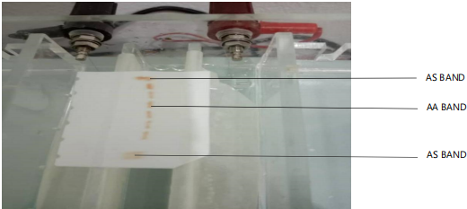
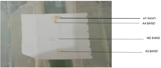
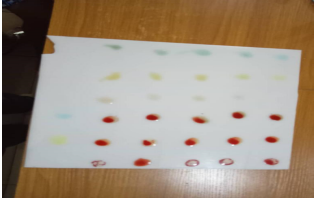
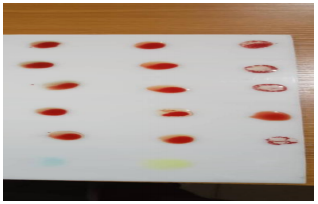
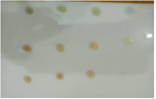
Figure 1 shows an electropherograms of the control samples showing AS band compared to subject samples which were all AA bands. Figure 2 is a representation of control blood, subject blood and mosquito blood meal samples. The blood samples exhibited the usual AS and AA bands; however, no band was observed for the mosquito blood meal. Figure 3 shows the blood group of the subjects and that of mosquito blood meal specimens, whereas that of Fig. 4 was strictly blood samples. Figure 5 shows agglutination in engorged mosquito samples and no agglutination of un-engorged mosquito's samples and water (control treatment). Below is the pictorial of the electrophoregrams of the study.
|
|
|
|
|
DISCUSSION
The identification of mosquito blood meal is a valuable tool in forensic investigations. It could offer vital evidence that links individuals to crime scenes, assists in determining when events occurred and even aids in identifying victims or suspects23-25. This study indicated that the use of cellulose acetate paper electrophoretic method in the identification and possible individualization of mosquito blood meal is still remote and laddered with difficulties in interpretation (Fig. 1-2). The results aligned with previous research by de Souza et al.17, cellulose acetate electrophoresis could not differentiate mosquito blood meals from the subject exposed to the mosquito bite. The unreliability of Hb genotype identification has spurred the inroads into advanced techniques. This could be the basis for the increasing applications of molecular techniques and other advanced techniques in the identification and further individualization of mosquito blood meals for various scientific utility26,27.
In furtherance to the identification and possible speciation of mosquito blood meal a serological approach was also utilized in this study. The findings established the possibility of identifying mosquito blood meal using ABO and Rhesus D antibodies, but that of individualization was invalid (Figure 3-5). The engorged mosquito blood meal exhibited an agglutination phenomenon irrespective of the type of blood groups, whereas the non-engorged did not agglutinate. This finding could be utilized in the identification of the presence of blood especially in crime scenes where blood could not be found and there is a denial of the presence of an individual. This study agreed with that of Rabelo et al.27, who reported the possibility of analyzing human blood found in hematophagous mosquitoes as biological trace evidence and with the findings of Santos et al.28 which indicates the presence of human blood.
Similarly, the finding also concurred with that of Tempelis and Lofy29, some of the earliest callers to this course of discussion with different animal species. They reported the use of anti-sera gotten from chickens for testing the frozen abdomens of engorged mosquitoes in gelatine capsules.
CONCLUSION
The study was able to establish the suitability of ABO and Rhesus D anti-sera in the identification of the presence of human blood in a mosquito engorged with blood meal. This could be of benefit in cases of denial of the presence of suspects at a crime scene. The study called for advanced molecular and biochemical approaches in the identification and individualization of mosquito blood meal.
SIGNIFICANCE STATEMENT
The application of mosquito blood meals in identifying and individualizing a suspect of a crime is quite expensive and scientifically clumsy. The study utilized simple techniques such as serology and Hb genotypes in making inroads into suspects’ exclusion and inclusion in a crime scene. The findings revealed that serology could be employed in identifying mosquito blood meal and by extension indicating the presence of someone within the crime scene
ACKNOWLEDGMENT
This research was supported by the 2021 TET Fund Institution-Based Research (IBR) Intervention granted to Dr. Eni-yimini Solomon Agoro (Ref:FUO/AP/TETF/21/IBR/001) of the Department of Biochemistry, Faculty of Science, Federal University Otuoke, Bayelsa State, Nigeria.
REFERENCES
- Soares, I.M.N., J.C. Polonio, J.A.C. Zequi and H.C. Golias, 2022. Molecular techniques for the taxonomy of Aedes Meigen, 1818 (Culicidae: Aedini): A review of studies from 2010 to 2021. Acta Trop., 236.
- Mullen, G.R. and L.A. Durden, 2009. Medical and Veterinary Entomology. 2nd Edn., Academic Press, New York, ISBN: 9780080919690, Pages: 637.
- Harbach, R.E., 2007. The Culicidae (Diptera): A review of taxonomy, classification and phylogeny. Zootaxa, 1668: 591-638.
- Phasomkusolsil, S., K. Pantuwattana, J. Tawong, W. Khongtak and Y. Kertmanee et al., 2015. The relationship between wing length, blood meal volume, and fecundity for seven colonies of Anopheles species housed at the Armed Forces Research Institute of Medical Sciences, Bangkok, Thailand. Acta Trop., 152: 220-227.
- Reeves, L.E., C.J. Holderman, E.M. Blosser, J.L. Gillett-Kaufman, A.Y. Kawahara, P.E. Kaufman and N.D. Burkett-Cadena, 2018. Identification of Uranotaenia sapphirina as a specialist of annelids broadens known mosquito host use patterns. Commun. Biol., 1.
- Toma, T., I. Miyagi and M. Tamashiro, 2014. Blood meal identification and feeding habits of Uranotaenia species collected in the Ryukyu Archipelago. J. Am. Mosq. Control Assoc., 30: 215-218.
- Forattini, O.P., A. de Castro Gomes, D. Natal, I. Kakitani and D. Marucci, 1987. Feeding preferences of Culicidae mosquitoes in the Ribeira Valley, S.Paulo State, Brazil. Rev. Saúde Pública, 21: 171-187.
- Damelio, R. and R.M. Gardner, 2001. Bloodstain Pattern Analysis: Its Function and a Historical Perspective. In: Bloodstain Pattern Analysis: With an Introduction to Crime Scene Reconstruction, Bevel, T. and R.M. Gardner (Eds.), CRC Press, Boca Raton, Florida, ISBN: 9780429247989, pp: 1-20.
- Curic, G., R. Hercog, Z. Vrselja and J. Wagner, 2014. Identification of person and quantification of human DNA recovered from mosquitoes (Culicidae). Forensic Sci. Int.: Genet., 8: 109-112.
- Attardo, G.M., I.A. Hansen and A.S. Raikhel, 2005. Nutritional regulation of vitellogenesis in mosquitoes: Implications for anautogeny. Insect Biochem. Mol. Biol., 35: 661-675.
- Wang, L., X. Lv, Y. Zhai, S. Fu and D. Wang et al., 2012. Genomic characterization of a novel virus of the family Tymoviridae isolated from mosquitoes. PLoS ONE, 7.
- Chadee, D.D. and J.C. Beier, 1997. Factors influencing the duration of blood-feeding by laboratory-reared and wild Aedes aegypti (Diptera: Culicidae) from Trinidad, West Indies. Ann. Trop. Med. Parasitol., 91: 199-207.
- Delatte, H., A. Desvars, A. Bouétard, S. Bord, G. Gimonneau, G. Vourc'h and D. Fontenille, 2010. Blood-feeding behavior ofAedes albopictus, a vector of chikungunya on La Réunion. Vector-Borne Zoonotic Dis., 10: 249-258.
- Washino, R.K. and J.G. Else, 1972. Identification of blood meals of hematophagous arthropods by the hemoglobin crystallization method. Am. J. Trop. Med. Hyg., 21: 120-122.
- McKinney, R.M., J.T. Spillane and P. Holden, 1972. Mosquito blood meals: Identification by a fluorescent antibody method. Am. J. Trop. Med. Hyg., 21: 999-1003.
- Kent, R.J., 2009. Molecular methods for arthropod bloodmeal identification and applications to ecological and vector-borne disease studies. Mol. Ecol. Resour., 9: 4-18.
- de Souza, S.S., A.R. Gomes, A.T.B. Guimarães, A.S. de Lima Rodrigues and L.P. de Matos et al., 2023. Cellulose microcrystalline: A promising ecofriendly approach to control Culex quinquefasciatus larvae. Sci. Total Environ., 901.
- Pierce, B.A., 2020. Genetics: A Conceptual Approach. 7th Edn., Macmillan Learning, New York, ISBN: 9781319216801, Pages: 944.
- Boorman, J., P.S. Mellor, P.F.L. Boreham and R.S. Hewett, 2009. A latex agglutination test for the identification of blood-meals of Culicoides (Diptera: Ceratopogonidae). Bull. Entomol. Res., 67: 305-311.
- Bryan, J.H. and M.E. Smalley, 1978. The use of ABO blood groups as markers for mosquito biting studies. Trans. R. Soc. Trop. Med. Hyg., 72: 357-360.
- Gunathilaka, N., T. Denipitiya, M. Hapugoda, W. Abeyewickreme and R. Wickremasinghe, 2016. Determination of the foraging behaviour and blood meal source of malaria vector mosquitoes in Trincomalee District of Sri Lanka using a multiplex real time polymerase chain reaction assay. Malar. J., 15.
- Agoro, E.Y.S. and O.B. Ilesanmi, 2022. Transference capacity of some heavy metals in cultivation-consumption chain of plantain harvested from Awka Etiti Anambra State. Asia Pac. J. Med. Toxicol., 11: 19-24.
- Gray, S.L., T.M. Tiedge, J.M. Butkus, T.J. Earp, S.E. Lindner and R. Roy, 2020. Determination of human identity from Anopheles stephensi mosquito blood meals using direct amplification and massively parallel sequencing. Forensic Sci. Int.: Genet., 48.
- Keven, J.B., M. Katusele, R. Vinit, D. Rodríguez-Rodríguez and M.W. Hetzel et al., 2021. Nonrandom selection and multiple blood feeding of human hosts by Anopheles vectors: Implications for malaria transmission in Papua New Guineaa. Am. J. Trop. Med. Hyg., 105: 1747-1758.
- Keller, J.I., B.A. Ballif, R.M.S. Clair, J.J. Vincent, M.C. Monroy and L. Stevens, 2017. Chagas disease vector blood meal sources identified by protein mass spectrometry. PLoS ONE, 12.
- Altahir, O., H. AbdElbagi, M. Abubakr, E.E. Siddig, A. Ahmed and N.S. Mohamed, 2022. Blood meal profile and positivity rate with malaria parasites among different malaria vectors in Sudan. Malar. J., 21.
- Rabelo, K.C.N., C.M.R. Albuquerque, V.B. Tavares, S.M. Santos and C.A. Souza et al., 2015. Trace samples of human blood in mosquitoes as a forensic investigation tool. Genet. Mol. Res., 14: 14847-14856.
- Santos, C.S., M.R. Pie, T.C. da Rocha and M.A. Navarro-Silva, 2019. Molecular identification of blood meals in mosquitoes (Diptera, Culicidae) in urban and forested habitats in Southern Brazil. PLoS ONE, 14.
- Tempelis, C.H. and M.F. Lofy, 1963. A modified precipitin method for identification of mosquito blood-meals. Am. J. Trop. Med. Hyg., 12: 825-831.
How to Cite this paper?
APA-7 Style
Solomon,
A.E., Teddy,
A.C., German,
I.C. (2024). Use of Anopheles Mosquito Bloodmeal’s Serology and Haemoglobin Genotypes in Crime Scene Forensics. Asian Journal of Biological Sciences, 17(4), 574-581. https://doi.org/10.3923/ajbs.2024.574.581
ACS Style
Solomon,
A.E.; Teddy,
A.C.; German,
I.C. Use of Anopheles Mosquito Bloodmeal’s Serology and Haemoglobin Genotypes in Crime Scene Forensics. Asian J. Biol. Sci 2024, 17, 574-581. https://doi.org/10.3923/ajbs.2024.574.581
AMA Style
Solomon
AE, Teddy
AC, German
IC. Use of Anopheles Mosquito Bloodmeal’s Serology and Haemoglobin Genotypes in Crime Scene Forensics. Asian Journal of Biological Sciences. 2024; 17(4): 574-581. https://doi.org/10.3923/ajbs.2024.574.581
Chicago/Turabian Style
Solomon, Agoro,, Eni-Yimini, Adias, Charles Teddy, and Ikimi, Charlse German.
2024. "Use of Anopheles Mosquito Bloodmeal’s Serology and Haemoglobin Genotypes in Crime Scene Forensics" Asian Journal of Biological Sciences 17, no. 4: 574-581. https://doi.org/10.3923/ajbs.2024.574.581

This work is licensed under a Creative Commons Attribution 4.0 International License.