Sanative Effect of Newly Developed Herbal Formulation SS-IM-21 Upon Ethanol Induced Hepatic Oxidative Stress Against Mice
-
Soumendra Darbar


Research and Development Division, Dey’s Medical Stores (Manufacturing) Limited, 62, Bondel Road, Kolkata 700019, West Bengal, India
Srimoyee SahaFaculty of Science, Jadavpur University, 188, Raja Subodh Chandra Mallick Road, Kolkata 700032, West Bengal, India
Atiskumar ChattopadhyayFaculty Council of Science, Jadavpur University, 188, Raja Subodh Chandra Mallick Road, Kolkata 700032, West Bengal, India
| Received 08 Dec, 2022 |
Accepted 29 Mar, 2023 |
Published 30 Jun, 2023 |
Background and Objective: Oxidative stress represents an imbalance between oxidant and antioxidant agents. Reactive oxygen species (ROS) derived from oxygen and nitrogen play a vital role in the propagation of liver injury by damaging cell membranes, micro and macromolecules. The objective of the study of this study was to find out the probable ameliorative effect of the newly developed herbal formulation (SS-IM-21) on serum and hepatic antioxidant enzymes activities in ethanol-induced oxidative stress in mice. Materials and Methods: Forty adult Swiss albino mice were randomly chosen and divided into four groups, Group-I as control, Group II ethanol treatment orally (50% v/v), Group III pre-treated with herbal formulation (SS-IM-21) with ethanol (50% v/v) and Group-IV only treated with herbal formulation (SS-IM-21) without ethanol daily. After the 6 weeks of the experimental period, the animals were euthanized and the livers were immediately removed and collected, kept frozen until analysis. Before the mice were killed blood samples were also collected to measure superoxide dismutase (SOD), catalase (CAT), glutathione peroxidase (GPx) and glutathione-S transferase (GST) activities in sera. Results: Major antioxidant enzymes like superoxide dismutase (SOD), catalase (CAT), glutathione peroxidase (GPx) and glutathione-S transferase (GST) activities in serum and liver significantly declined in the ethanol-treated mice to control. Treatment with herbal formulation (SS-IM-21) suppresses oxidative stress in comparison with the ethanol group. Conclusion: In the current study the results established that treatment with herbal formulation (SS-IM-21) might be a potent antioxidant that exerts beneficial effects on both superoxide dismutase (SOD), catalase (CAT), glutathione peroxidase (GPx) and glutathione-S transferase (GST) activities in ethanol intoxicated mice and inhibit organ damage.
| Copyright © 2023 Darbar et al. This is an open-access article distributed under the Creative Commons Attribution License, which permits unrestricted use, distribution, and reproduction in any medium, provided the original work is properly cited. |
INTRODUCTION
Inflammatory responses are generally developed by the involvement of reactive oxygen species (ROS), thereby affecting normal cellular physiology and playing a significant role in pathological conditions1. The free radicals, apart from being involved in damaging cellular components, do play a significant role in ethanol-induced organ toxicity2. The liver is the main organ where the metabolism of xenobiotics to a large extent takes place. Most of the time by-products of such metabolism make severe toxic effects and produce cellular imbalance3. This could lead to liver damage and the emergence of hepatic disorders. In very frequent oxygen-containing by-product molecules damage liver cells through oxidation. They produce oxidative stress and generate an enormous amount of free radicals which affects cell survival because of membrane damage through the oxidative damage of lipid, protein and irreversible DNA modification4. This condition destroys the balance between the production of reactive oxygen species (ROS) and the ability of the body to counteract or detoxify their harmful effects through neutralization by antioxidants5. Excessive formation and insufficient removal of free radicals lead to destructive and irreversible cell damage. A scientific study reviled that oxidative damage is aggravated by the decrease in various antioxidant enzymes activities such as superoxide dismutase, catalase (CAT), glutathione S-transferase (GST) and glutathione peroxidase (GPx) which acts as a free radical scavenger in conditions associated with oxidative stress6-8.
Herbal drugs, used in Indian systems of medicine are however claimed to be effective and safe in such ailments. These drugs are considered benign and are of particular value in the treatment of chronic diseases requiring prolonged therapy. Plant medicines are more often used in combination rather than in a single to get the maximum benefit from their combined strength. The SS-IM-21, a multi-herbal formulation expected very useful as natural hepatoprotective medicine, which compose of several three Indian medicinal plants such as Andrographis paniculata, Withania somnifera and Ocimum sanctum9-11. Because of this, the present study aimed to investigate whether the oral administration of a newly developed herbal formulation (SS-IM-21) at a dose of 200 mg kg–1 of the body weight can control the hepatic and renal oxidative stress caused by ethanol intoxication in male mice and protect the cell from redox damage.
MATERIALS AND METHODS
Study area: The study was conducted in the Pharmacology Laboratory of Jadavpur University, Kolkata, India. It was carried out in the last quarter of 2020 and the first quarter of 2021.
Drugs and chemicals: All the medicinal plants used in this experiment were collected from a registered supplier. Ethanol and phosphate buffer were obtained from Merck, India. Antioxidant enzyme study kits were purchased from E-Mark Germany. Other chemicals were obtained from local sources and were of analytical grade.
Animals: In this study, 40 young Swiss albino mice weighing 25-27 g have been randomly included for the study. The animals have been housed in healthy atmospheric conditions, with normal feeding, drinking and medical care based on the CPCSEA guidelines. Before dosing, they were acclimatized for 7 days to light from 06:00 to 18:00 hrs, alternating with 12 hrs darkness. The animals were housed in stainless steel cages in an air-conditioned room with a temperature maintained at 25±2°C. Mice were allowed a standard chow diet (Amrut feeds, Pranav Agro, New Delhi, India) throughout the experiment and water ad libitum.
Ethical consideration: The experimental procedures were approved by the Institutional Animal Ethics Committee (IAEC) (Approval No. 15/IAEC/Dey’s/s/2016).
Experimental design: Adult healthy mice were divided randomly into four experimental groups, each consisting of ten mice, that were treated as follows: Group I received the vehicle and served as a control, Group II received Ethanol (50% v/v), Group III received Ethanol (50% v/v) along with Herbal formulation (SS-IM-21) (200 mg kg–1) and Group IV received only Herbal formulation (SS-IM-21) (200 mg kg–1) for 6 weeks.
Serum preparation and organ collection: After complications of the experimental period, blood was collected from the retro-orbital plexus. Collected blood samples stay for 1 hr 15 min at room temperature and then centrifuged at 6500 rpm for 15 min to obtain clear serum. The serum was stored in aliquots at -70°C till used for the estimation of various antioxidant enzymes.
After proper euthanasia liver was removed, washed three times in ice-cold saline and blotted individually on ash-free filter paper, used for the preparation of tissue homogenates for estimation of tissue SOD, CAT, GSH and GST activities.
Tissue homogenate preparation: A small portion of liver tissues was weighed and homogenized separately with a Potter-Elvenhjem tissue homogenizer in phosphate buffer saline (PBS) at 50 mM pH (7.4) for estimation of protein content, SOD, CAT, GST and CAT enzymes activities.
Determination of lipid peroxidation: The process of lipid peroxidation (LPO) measurement was carried out using a lipid peroxidation (MDA) assay kit (Sigma-Aldrich Ltd., UK) following the manufacturer’s instructions. In this assay, lipid peroxidation is determined by the reaction of malondialdehyde (MDA) with thiobarbituric acid (TBA) to form a colorimetric product, proportional to the MDA present. To form the MDA-TBA adduct, the TBA solution (600 mL) was added to each sample and incubated at 95°C for 60 min, before cooling to room temperature in an ice bath for 10 min. Each reaction mixture (200 mL) was transferred into a 96-well plate for analysis. The absorbance was measured by UV-spectrophotometer (Shimadzu UV-1280) at 532 nm12.
Assay of antioxidant enzyme activities
Determination of catalase (CAT) activity: The CAT activity was determined by the method of Ball et al.13. In brief, a quartz cuvette, phosphate buffer (pH 7.0) and a sample were added and the reaction was started by the addition of H2O2. The decomposition of H2O2 was monitored at 240 nm.
Determination of superoxide dismutase (SOD) activity: The SOD activity in serum, liver and kidney homogenate was assessed according to the method of Chandran et al.14 with slight modification. In a quartz cuvette, 1 mL ois-HCl buffer, containing diethylene triaminopentaacetic acid and pyrogallol was mixed with 20 μL of kidney samples. The difference in absorbance was measured at 440 nm.
Determination of reduced glutathione (GSH) activity: Reduced glutathione (GSH) was determined from liver homogenate spectrophotometrically according to Sánchez-Illana et al.15. The decrease in absorbance was determined at 340 nm.
Determination of glutathione-S transferase (GST) activity: The GST activity of serum, liver and kidney tissues was investigated by the method of El-Refaei et al.16 with slight modification. 1-chloro-2-4-di-nitrobenzene is neutralized by the enzyme in the presence of glutathione as a cosubstrate. The change in absorbance was measured at 340 nm.
Statistical analysis: All analyses were carried out using the SPSS software, version 20.0. A One-way Analysis of Variance (ANOVA, p<0.05) and Tukey’s test were used to determine significant differences between groups. The values were stated as Mean±SD.
RESULTS
Protective effect of herbal formulation (SS-IM-21) on serum and liver MDA content in ethanol toxicity: The effect of a herbal combination composed of Indian medicinal plants (Table 1) on serum and liver MDA levels was depicted in Table 2. Serum MDA content in the ethanol-intoxicated mice was significantly higher (106.39±1.59 vs 46.28±1.14) than that of the controls. Pre-treatment with Herbal formulation (SS-IM-21) (Table 1) significantly decreased (49.62±0.89 vs 106.39±1.59) serum MDA content compared with ethanol-intoxicated mice.
|
| Table 1: | Gross composition of solvent extracted processed soyabean based diets | |||
| Scientific name | Common name |
Family |
Quantity (mg) |
| Andrographis paniculata | Kalmegh |
Acanthaceae |
50 |
| Withania somnifera | Ashwagandha |
Solanaceae |
25 |
| Ocimum sanctum | Tulsi |
Lamiaceae |
50 |
| Each capsule contains: Powder and extractive derived from the above stated ingradients | |||
| Table 2: | Effect of newly developed herbal formulation (SS-IM-21) on serum and liver MDA levels in ethanol intoxicated mice | |||
MDA (nmol g–1) |
||
| Group | Serum |
Liver |
| Control | 46.28±1.14 |
32.61±1.16 |
| Ethanol (50% v/v) | 106.39±1.59# |
78.47±0.81# |
| Ethanol+SS-IM-21 (200 mg kg–1) | 49.62±0.89* |
38.06±0.81* |
| SS-IM-21 (200 mg kg–1) | 44.11±1.55* |
33.68±0.99* |
| Values are Mean±SD of ten observations, #Significant difference from control mice (p<0.001) and *Significant difference from ethanol intoxicated group (p<0.05) | ||
Hepatic MDA content in the ethanol-intoxicated mice was significantly elevated (78.47±0.81 vs 32.61±1.16) than that of controls. Pre-treatment with herbal formulation (SS-IM-21) (Table 1) significantly declined (38.06±0.81 vs 78.47±0.81) liver MDA content compared with ethanol-intoxicated mice.
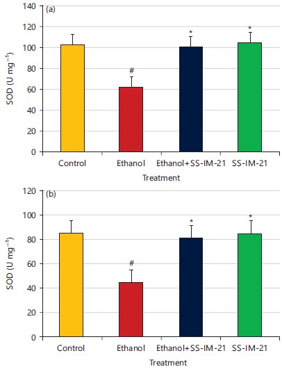
Protective effect of herbal formulation (SS-IM-21) on serum and liver SOD activity in ethanol toxicity: Serum and liver superoxide dismutase (SOD) activities are depicted in Fig. 1a-b. Serum SOD activity in the ethanol-intoxicated mice was significantly less (61.85±2.48 vs 102.64±3.98) than that of the controls. Pre-treatment with herbal formulation (SS-IM-21) (Table 1) significantly increased (100.69±3.02 vs 61.85±2.48) serum SOD activity compared with ethanol-intoxicated mice.

|
Hepatic superoxide dismutase (SOD) activity in the ethanol-intoxicated mice was significantly less (44.85±2.06 vs 85.27±1.09) than that of controls. Pre-treatment with herbal formulation (SS-IM-21) (Table 1) significantly increased (81.02±1.69 vs 44.85±2.06) hepatic SOD activity compared with ethanol-intoxicated mice.
Protective effect of herbal formulation (SS-IM-21) on serum and liver CAT activity in ethanol toxicity: Serum and liver catalase (CAT) activities are depicted in Fig. 2a-b. Serum CAT activity in the ethanol-intoxicated mice was significantly less (156.29±4.62 vs 225.17±5.02) than that of the controls. Pre-treatment with herbal formulation (SS-IM-21) (Table 1) significantly increased (202.05±3.67 vs 156.29±4.62) serum CAT activity compared with ethanol-intoxicated mice.
Hepatic catalase (CAT) activity in the ethanol-intoxicated mice was significantly less (101.26±2.57 vs 175.48±4.08) than that of controls. Pre-treatment with Herbal formulation (SS-IM-21) (Table 1) significantly increased (170.65±4.21 vs 101.26±2.57) hepatic CAT activity compared with ethanol-intoxicated mice.

|
Protective effect of herbal formulation (SS-IM-21) on serum and liver GSH activity in ethanol toxicity: Serum and liver-reduced glutathione (GSH) activities are depicted in Fig. 3a-b. Serum GSH activity in the ethanol-intoxicated mice was significantly less (19.62±0.92 vs 38.91±1.02) than that of the controls. Pre-treatment with herbal formulation (SS-IM-21) (Table 1) significantly increased (36.07±0.61 vs 19.62±0.92) serum GSH activity compared with ethanol-intoxicated mice.
Hepatic reduced glutathione (GSH) activity in the ethanol-intoxicated mice was significantly less (9.06±0.48 vs 19.62±0.24) than that of controls. Pre-treatment with herbal formulation (SS-IM-21) (Table 1) significantly increased (21.05±0.31 vs 9.06±0.48) renal GSH activity compared with ethanol-intoxicated mice.
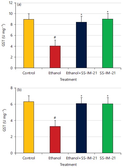
Protective effect of herbal formulation (SS-IM-21) on serum and liver glutathione-S transferase (GST) activity in ethanol toxicity: Serum, kidney and liver glutathione-S transferase (GST) activities are depicted in Fig. 4a-b. Serum GSH activity in the ethanol-intoxicated mice was significantly less (4.11±0.26 vs 8.92±0.34) than that of the controls. Pre-treatment with herbal formulation (SS-IM-21) (Table 1) significantly increased (8.47±0.45 vs 4.11±0.26) serum GSH activity compared with ethanol-intoxicated mice.
|
Hepatic glutathione-S transferase (GST) activity in the ethanol-intoxicated mice was significantly less (3.28±0.11 vs 6.35±0.24) than that of controls. Pre-treatment with herbal formulation (SS-IM-21) (Table 1) significantly increased (6.11±0.26 vs 3.28±0.11) renal GSH activity compared with ethanol-intoxicated mice.
DISCUSSION
The newly developed medicinal plant combination SS-IM-21 produced an ameliorative effect against ethanol-induced oxidative stress17. The combination controls the liver function enzyme levels and maintains serum protein concentration in the preclinical model. This combination effectively sounds against liver fibrosis due to its synergistic function. The previous study showed that herbs and herbal formulation in different concentration is beneficial to maintain normal hepatic function18,19. This medicine is probably very helpful to maintain liver oxidative stress by controlling oxidative enzymes20-25. Consumption of ethanol produces reactive oxygen species (ROS) in every mammalian cell. The liver and kidney are the two target organs for oxidative stress during ethanol intoxication. Various free radicals such as Superoxide anion (O2•–), Hydroxyl radical (OH•) and Hydrogen peroxide (H2O2) are the major ROS generated during normal redox reactions in our body that produce cytotoxic effects. They are generally neutralized by the defensive action of the endogenous antioxidant system, primarily composed of glutathione17, superoxide dismutase18, glutathione peroxidase and catalase. The imbalance between the generation and neutralization of ROS can create severe oxidative stress-induced damage, consequently, ROS accumulation may cause protein oxidation leading to the disruption of cell membranes, organelles and loss of function21.
Lipid peroxidation is commonly used as a marker for the induction of oxidative stress in cells. The level of MDA, which is generated as an end product during the oxidation of lipids, was used as a marker of lipid peroxidation22-25. Treatment of mice with ethanol (50% v/v) increased lipid peroxidation as shown by elevated MDA levels in serum, liver and kidney tissues. This situation suggests the induction of oxidative stress in cells. The application of multi-herbal formulation (SS-IM-21) significantly reduced the serum MDA level. The treatment also reduced the hepatic and renal MDA levels which indicates that this herbal medicine maintains the normal fluidity of the cell membrane which plays a vital role in cell functioning.
Superoxide dismutase (SOD), catalase (CAT), glutathione (GSH) and glutathione-S transferase (GST) are the most common antioxidant enzymes inhibited by ethanol intoxication during oxidative damage. In the present study, we aim to determine the possible therapeutic effect of herbal formulation (SS-IM-21) upon SOD, GPx, GST and CAT enzyme activities as an indicator of oxidative stress. A scientific study reviled that SOD is an enzyme against the superoxide radical and catalyzes its dismutation into H2O2, which is utilized by CAT or GPx26-30. On the other hand, GST catalyzes the conjugation of several substrates to the thiol group of glutathione, transforming toxic materials into less toxic forms31-33. In the present study, oral administration of ethanol (50% v/v) on mice significantly reduced the antioxidant enzyme activities as compared to control untreated animals which supported the previous experiment that chronic consumption of ethanol generates free radicals which reduced the antioxidant enzyme activities. The generation of reactive oxygen species (ROS) within the cell decreased cellular performance by changing the antioxidant enzyme’s actions. Treatment with multi-herbal formulation (SS-IM-21) at a dose of 200 mg/kg/day on mice who are intoxicated with ethanol, significantly elevated the SOD, GPx, GST and CAT enzyme activities. This herbal formulation (SS-IM-21) inhibits the free radical production within the cell which indicated that the synergistic action of various plant compounds in a single medicine may be potent to prevent cellular oxidative stress and boost the cell their normal function.
CONCLUSION
Chronic consumption of ethanol is accompanied by increased serum, hepatic and renal tissue oxidative stress, which is characterized by a reduction in the antioxidant enzyme activities and glutathione levels that correlate with the increase in MDA in the tissues. This may probably contribute to the additional progression of ethanol intoxication-related problems. Treatment with multi-herbal formulation (SS-IM-21) normalized the serum and tissues’ antioxidant enzyme activities by suppressing extensive ROS generation during ethanol intoxication. So, a newly developed multi-herbal formulation (SS-IM-21) composed of medicinal herbs may be a potent drug that is sound for the prevention of cellular oxidative stress.
SIGNIFICANCE STATEMENT
This study discovered the newly developed multi-herbal formulation (SS-IM-21) composed of three essential Indian medicinal plants namely Andrographis paniculata, Withania somnifera, Ocimum and Sanctum the first time in the Indian traditional system of medicine that can be beneficial for the treatment of various liver complications like oxidative stress. This study will help the researchers to uncover the critical areas of the molecular mechanism of the drug action and efficacy of the multi-herbal formulation that many researchers were not able to explore. Thus a new theory on herbal formulation is safe and non-toxic and very useful for chronic liver disease.
ACKNOWLEDGMENTS
The authors are thankful to Professor (Dr.) T.K. Pal, Department of Pharmaceutical Technology, Jadavpur University, Kolkata-700032 and Professor S.K. Pal, Senior Professor Department of Chemical, Biological and Macromolecular Sciences, S.N. Bose National Centre for Basic Sciences, JD Block, Sector III, Salt Lake City, Kolkata for their valuable suggestions and Mr. Gautam Dey, M.D. and Mr. Ranajit Dey, Jt. M.D. for facilities and encouragement during this investigation.
REFERENCES
- Goc, Z., E. Kapusta, G. Formicki, M. Martiniaková and R. Omelka, 2019. Effect of taurine on ethanol-induced oxidative stress in mouse liver and kidney. Chin. J. Physiol., 62: 148-156.
- Jing, L., C.M. Jin, S.S. Li, F.M. Zhang and L. Yuan et al., 2012. Chronic alcohol intake-induced oxidative stress and apoptosis: Role of CYP2E1 and calpain-1 in alcoholic cardiomyopathy. Mol. Cell Biochem., 359: 283-292.
- Simplicio, J.A., G.T. do Vale, N.A. Gonzaga, L.N. Leite and U.V. Hipólito et al., 2017. Reactive oxygen species derived from NAD(P)H oxidase play a role on ethanol-induced hypertension and endothelial dysfunction in rat resistance arteries. J. Physiol. Biochem., 73: 5-16.
- Darbar, S., M.R. Chakraborty, S. Chattarjee and B. Ghosh, 2009. Protective effect of livina, a polyherbal liquid formulation against ethanol induced liver damage in rats. Ancient Sci. Life, 28: 14-17.
- Khodaei, F., H. Kholghipour, M. Hosseinzadeh and M. Rashedinia, 2019. Effect of sodium benzoate on liver and kidney lipid peroxidation and antioxidant enzymes in mice. J. Rep. Pharm. Sci., 8: 217-223.
- El-Shenawy, N.S., B. El-Ahmary and R.A. Al-Eisa, 2011. Mitigating effect of ginger against oxidative stress induced by atrazine herbicides in mice liver and kidney. J. Agric. Sci. Food Res., 2: 1000107.
- Darbar, S., S. Saha, K. Pramanik and A. Chattopadhyay, 2021. Antioxidant and immunomodulatory effect of AKSS16-LIV01-a multi herbal formulation against ethanol induced liver dysfunction in mice. Clin. Phytosci., 7: 80.
- Cederbaum, A.I., Y. Lu and D. Wu, 2009. Role of oxidative stress in alcohol-induced liver injury. Arch. Toxicol., 83: 519-548.
- Darbar, S. and S. Chattopadhyay, 2018. Single dose acute oral toxicity of livina, a polyherbal formulation in mice model. Eur. J. Pharm. Med. Res., 5: 492-495.
- Darbar, S., S. Saha, S. Chattopadhyay and A. Chattapadhyay, 2020. Anti-stress activity (in-vivo) of multi herbal capsule-Trasina® in experimental murine model. Asian J. Pharm. Res. Dev., 8: 52-58.
- Darbar, S., S. Saha, K. Pramanik and A. Chattopadhyay, 2018. Preliminary acute oral toxicity study of a newly developed herbal formulation. World J. Pharm. Res., 7: 924-930.
- Devi, G.S., M.H. Prasad, I. Saraswathi, D. Raghu, D.N. Rao and P.P. Reddy, 2000. Free radicals antioxidant enzymes and lipid peroxidation in different types of leukemias. Clin. Chim. Acta, 293: 53-62.
- Ball, B.A., C.G. Gravance, V. Medina, J. Baumber and I.K.M. Liu, 2000. Catalase activity in equine semen. Am. J. Vet. Res., 61: 1026-1030.
- Chandran, R., A.A. Sivakumar, S. Mohandass and M. Aruchami, 2005. Effect of cadmium and zinc on antioxidant enzyme activity in the gastropod, Achatina fulica. Comp. Biochem. Phys. C.: Toxicol. Pharmacol., 140: 422-426.
- Sánchez-Illana, Á., F. Mayr, D. Cuesta-García, J.D. Piñeiro-Ramos and A. Cantarero et al., 2018. On-capillary surface-enhanced Raman spectroscopy: Determination of glutathione in whole blood microsamples. Anal. Chem., 90: 9093-9100.
- El-Refaei, M.F., and E.A.A. Abdallah, 2021. Protective effects of caffeic acid phenethyl ester on cadmium-induced testicular injury: A crucial role of antioxidant enzymes in male mice infertility. Heliyon, 7: e06965 .
- Hsu, J.Y. , H.H. Lin, C.C. Hsu, B.C. Chen and J.H. Chen, 2018. Aqueous extract of pepino (Solanum muriactum Ait) leaves ameliorate lipid accumulation and oxidative stress in alcoholic fatty liver disease. Nutrients, 10: 931.
- Shi, Y., L. Zhong, Y. Fan, J. Zhang and H. Zhong et al., 2022. The protective effect of mulberry leaf flavonoids on high-carbohydrate-induced liver oxidative stress, inflammatory response and intestinal microbiota disturbance in Monopterus albus. Antioxidants, 11: 976.
- Sun, Y., S. Cai, Y. Zhang, N. Ma, J. Yi, X. Hu and T. Wang, 2022. Protective effect of Rhus chinensis Mill. fruits on 3,5-diethoxycarbonyl-1,4-dihydrocollidine-induced cholestasis in mice via ameliorating oxidative stress and inflammation. Nutrients, 14: 4090.
- Li, Y., W. Cai, Z. Ai, C. Xue, R. Cao and N. Dong, 2023. Protective effects of sinomenine hydrochloride on lead-induced oxidative stress, inflammation, and apoptosis in mouse liver. Environ. Sci. Pollut. Res., 30: 7510-7521.
- Abdelmagid, A.D., A.M. Said, E.A. Abd El-Gawad, S.A. Shalaby and M.A.O. Dawood, 2022. Glyphosate-induced liver and kidney dysfunction, oxidative stress, immunosuppression in Nile tilapia, but ginger showed a protection role. Vet. Res. Commun.
- Figueredo, K.C., C.G. Guex, A.R.H. da Silva, C.L. Lhamas and A.M. Engelmann et al., 2022. In silico and in vivo protective effect of Morus nigra leaves on oxidative damage induced by iron overload. Drug Chem. Toxicol., 45: 2814-2824
- Cox, F.F., A. Misiou, A. Vierkant, N. Ale-Agha, M. Grandoch, J. Haendeler and J. Altschmied, 2022. Protective effects of curcumin in cardiovascular diseases-impact on oxidative stress and mitochondria. Cells, 11: 342.
- Luo, T., S. Jiang, B. Zhou, Q. Song and J. Du et al., 2022. Protective effect of isoorientin on oleic acid-induced oxidative damage and steatosis in rat liver cells. Front. Pharmacol., 13: 818159.
- Mohamed, E.E., O.M. Ahmed, A. Abdel-Moneim, K.M.A. Zoheir and B.H. Elesawy et al., 2022. Protective effects of naringin-dextrin nanoformula against chemically induced hepatocellular carcinoma in Wistar rats: Roles of oxidative stress, inflammation, cell apoptosis, and proliferation. Pharmaceuticals, 15: 1558.
- Harris, P.S., S.R. Roy, C. Coughlan, D.J. Orlicky and Y. Liang et al., 2015. Chronic ethanol consumption induces mitochondrial protein acetylation and oxidative stress in the kidney. Redox Biol., 6: 33-40.
- Milat, A.M., I. Mudnić, I. Grković, N. Ključević and M. Grga et al., 2017. Effects of white wine consumption on weight in rats: Do polyphenols matter? Oxid. Med. Cell. Longevity, 2017: 8315803.
- Rehm, J., C. Mathers, S. Popova, M. Thavorncharoensap, Y. Teerawattananon and J. Patra, 2009. Global burden of disease and injury and economic cost attributable to alcohol use and alcohol-use disorders. Lancet, 373: 2223-2233.
- Rossi, R.E., D. Conte and S. Massironi, 2015. Diagnosis and treatment of nutritional deficiencies in alcoholic liver disease: Overview of available evidence and open issues. Digestive Liver Dis., 47: 819-825.
- Abid, Z.B., M. Feki, A. Hedhili and M.H. Hamdaoui, 2007. Artemisia herba-alba Asso (Asteraceae) has equivalent effects to green and black tea decoctions on antioxidant processes and some metabolic parameters in rats. Ann. Nutr. Metab., 51: 216-222.
- Zararsiz, I., M. Sarsilmaz, U. Tas, I. Kus, S. Meydan and E. Ozan, 2007. Protective effect of melatonin against formaldehyde-induced kidney damage in rats. Toxicol. Ind. Health, 23: 573-579.
- Kankofer, M., G. Kolm, J. Aurich and C. Aurich, 2005. Activity of glutathione peroxidase, superoxide dismutase and catalase and lipid peroxidation intensity in stallion semen during storage at 5°C. Theriogenology, 63: 1354-1365.
- Abuja, P.M. and R. Albertini, 2001. Methods for monitoring oxidative stress, lipid peroxidation and oxidation resistance of lipoproteins. Clin. Chim. Acta, 306: 1-17.
How to Cite this paper?
APA-7 Style
Darbar,
S., Saha,
S., Chattopadhyay,
A. (2023). Sanative Effect of Newly Developed Herbal Formulation SS-IM-21 Upon Ethanol Induced Hepatic Oxidative Stress Against Mice. Asian Journal of Biological Sciences, 16(2), 145-154. https://doi.org/10.3923/ajbs.2023.145.154
ACS Style
Darbar,
S.; Saha,
S.; Chattopadhyay,
A. Sanative Effect of Newly Developed Herbal Formulation SS-IM-21 Upon Ethanol Induced Hepatic Oxidative Stress Against Mice. Asian J. Biol. Sci 2023, 16, 145-154. https://doi.org/10.3923/ajbs.2023.145.154
AMA Style
Darbar
S, Saha
S, Chattopadhyay
A. Sanative Effect of Newly Developed Herbal Formulation SS-IM-21 Upon Ethanol Induced Hepatic Oxidative Stress Against Mice. Asian Journal of Biological Sciences. 2023; 16(2): 145-154. https://doi.org/10.3923/ajbs.2023.145.154
Chicago/Turabian Style
Darbar, Soumendra, Srimoyee Saha, and Atiskumar Chattopadhyay.
2023. "Sanative Effect of Newly Developed Herbal Formulation SS-IM-21 Upon Ethanol Induced Hepatic Oxidative Stress Against Mice" Asian Journal of Biological Sciences 16, no. 2: 145-154. https://doi.org/10.3923/ajbs.2023.145.154

This work is licensed under a Creative Commons Attribution 4.0 International License.