
Effects of Ozone Therapy on Cardiac and Pulmonary Tissues in Cecal Perforation-Induced Sepsis in Streptozotocin-Induced Diabetic Mice
-
Bilgin Hasret Şimşek
Faculty of Medicine, Department of Anesthesiology and Reanimation, Gazi University, Ankara 06500, Türkiye
Hüseyin GöbütFaculty of Medicine, Department of Anesthesiology and Reanimation, Gazi University, Ankara 06500, Türkiye
Nurten İnanFaculty of Medicine, Department of General Surgery, Gazi University, Ankara 06500, Türkiye
Mustafa Arslan

Application and Research Centre for Life Sciences, Gazi University, Ankara 06830, Türkiye
Kürşat DikmenFaculty of Medicine, Department of General Surgery, Gazi University, Ankara 06500, Türkiye
Şaban Cem SezenKırıkkale University Faculty of Medicine, Department of Histology and Embryology, Kırıkkale 71450, Türkiye
Mustafa KavutçuFaculty of Medicine, Department of Medical Biochemistry, Gazi University, Ankara 06500, Türkiye
Ayşegül KüçükFaculty of Medicine, Department of Physiology, Bandırma Onyedi Eylül University, Bandırma 10250, Türkiye
| Received 18 Mar, 2026 |
Accepted 19 Oct, 2026 |
Published 31 Dec, 2026 |
Background and Objective: Sepsis is a life-threatening condition characterized by systemic inflammation and multi-organ damage, with the heart and lungs being particularly vulnerable. Diabetes exacerbates sepsis-induced oxidative stress and tissue injury, highlighting the need for effective adjunctive therapies such as ozone. This study evaluated the effects of ozone therapy as an adjuvant treatment on cardiac and pulmonary tissues in a diabetic mouse model of cecal perforation-induced sepsis. Materials and Methods: A total of 46 Swiss albino mice were used, with diabetes induced in 40 mice using streptozotocin (125 mg/kg, IP). After one month, mice were divided into control and experimental groups, including diabetes, ozone-treated, sepsis-induced, and combined treatment groups. Ozone (20 μg/mL, 1 mL, IP) was administered before or after Cecal Ligation and Perforation (CLP). After 24 hrs, heart and lung tissues were collected for histopathological and biochemical analyses. Results: Sepsis and diabetes significantly increased inflammatory and oxidative damage in cardiac and pulmonary tissues, as evidenced by elevated MDA levels and reduced CAT activity. Ozone therapy reduced tissue inflammation, decreased MDA levels, and increased CAT activity. Pre-treatment with ozone showed greater protective effects than post-treatment. Conclusion: Ozone therapy mitigates cardiac and pulmonary damage in diabetic sepsis, with pre-treatment providing more effective protection than post-treatment.
| Copyright © 2026 Şimşek et al. This is an open-access article distributed under the Creative Commons Attribution License, which permits unrestricted use, distribution, and reproduction in any medium, provided the original work is properly cited. |
INTRODUCTION
The global prevalence of diabetes mellitus (DM) has increased significantly over the past two decades and continues to rise. The number of cases has increased from an estimated 30 mL in 1985 to 463 mL in 2019, and it is projected that 642 mL individuals will be affected by diabetes by 20401. The underlying reasons include abnormalities in cell-mediated immunity and phagocyte function associated with hyperglycemia, as well as reduced vascularization. Hyperglycemia facilitates the colonization and growth of various microorganisms. In addition to many common infections occurring more frequently and severely in the diabetic population, some rare infections are observed exclusively in individuals with diabetes1-3. It is well established that in diabetes, the antioxidant defense system is insufficient to counteract oxidative stress resulting from the oxidation or peroxidation of lipids, proteins, and nucleic acids a process driven by hyperglycemia’s stimulation of reactive oxygen species production The increase in free radical production is one of the key factors contributing to the onset and progression of diabetic complications4.
Sepsis is a systemic infection associated with life-threatening organ dysfunction caused by a dysregulated host response to infection. Common clinical features include signs of infection along with organ dysfunctions (confusion, tachypnea, hypotension, and hepatic, renal, and hematologic impairments). Both pro-inflammatory and anti-inflammatory responses occur, accompanied by significant alterations in the pathways leading to organ dysfunction1,5,6. Organ dysfunction observed in sepsis and septic shock is associated with impaired production of high-energy ATP molecules within the mitochondria, resulting from oxidative damage to the electron transport chain and cytokine-mediated inhibition of the pyruvate dehydrogenase enzyme5. These reactions result in the production of free radicals with high oxidative potential, leading to lipid peroxidation. Factors such as lipid peroxidation have been associated with the development of multiple organ dysfunction7.
The current clinical approach in treatment begins with early diagnosis and identification of the source, with early antibiotic therapy playing a crucial role. In addition to antibiotics, steroids also play a significant role; however, it has been shown that steroids reduce mortality by only about 10%. Although antibiotics remain the cornerstone of sepsis treatment, global antibiotic resistance complicates management. Currently, antibiotic resistance is classified by the World Health Organization as one of the top ten global public health threats to humanity. Due to the difficulty and insufficiency in developing new antibacterial agents, alternative bactericidal therapies are highly desirable8.
The cardiovascular and respiratory systems are among the most frequently affected organ systems in sepsis. Sepsis-associated cardiovascular changes include vasoplegic shock, myocardial dysfunction, altered microvascular flow, and widespread endothelial injury. Sepsis is characterized by platelet and leukocyte adhesion, activation, increased expression of endothelial adhesion molecules, and activation of the coagulation cascade. This process results in widespread endothelial damage as well as shedding of the endothelial glycocalyx. Consequently, disproportionate vasodilation, increased vascular permeability, and microvascular thrombosis are further exacerbated9.
Another common complication of sepsis is acute lung injury (ALI). The ALI is a major cause of short-term mortality and reduced long-term quality of life. Acute lung injury (ALI) and its more severe form, acute respiratory distress syndrome (ARDS), may result from a variety of pathological processes that directly or indirectly affect the lungs. Approximately 40% of patients with sepsis develop ALI/ARDS10,11. In response to bacterial infection, neutrophil activation in the lungs leads to ALI, and the infiltration of polymorphonuclear neutrophils (PMNs) represents the primary mechanism of sepsis-induced pulmonary dysfunction and injury12. Protein-rich pulmonary edema caused by increased vascular permeability is the main pathological finding in the early stage of ALI/ARDS. Alveolar flooding and alveolar collapse (due to interstitial edema) result in decreased compliance and reduced functional residual capacity10,13,14. ARDS is characterized by the accumulation of fluid and cellular debris within alveoli that are normally air-filled, due to inflammatory mechanisms. Furthermore, fibrosis that occurs during the fibroproliferative phase of ARDS has been shown to cause loss of aeration and an increase in lung radiological density14.
Oxidative stress is recognized as an important component of various diseases. In these conditions, oxidative stress results from the action of free radicals and non-radical compounds. Free radicals are atoms or molecules with unpaired electrons that can be generated during physiological or pathological reactions15. The most well-known oxidative compounds are reactive oxygen species (ROS) and reactive nitrogen species (RNS)16.
Ozone (O3) is a gas consisting of three oxygen atoms in a cyclic structure. It induces antioxidant stress, enhances resistance to oxidative stress, and has been used as an antioxidant agent in inflammation. Oxygen-ozone therapy is known as a bio-oxidative procedure, and low-dose ozone administration has been shown to inhibit prostaglandin synthesis, the release of bradykinin and algogenic substances, and the secretion of proteinases from macrophages and polymorphonuclear leukocytes17,18. Ozone therapy is an emerging treatment strategy that exerts its effects through complex mechanisms such as antioxidant properties, immunomodulatory capacity, and modulation of local microcirculation19. The efficacy of oxygen ozone (O2-O3) therapy is thought to be partly related to the moderate oxidative stress generated by the interaction of O3 with biological components. O2-O3 therapy is widely used as an adjunctive therapeutic option in a variety of pathological conditions characterized by chronic inflammatory processes and immune hyperactivation20.
In diabetes, abnormalities in cell-mediated immunity, impaired phagocyte function associated with hyperglycemia, and increased oxidative stress due to hyperglycemia contribute to organ dysfunction and significantly increase mortality. Among sepsis-related organ failures, the heart and lungs are the two organs most critically affecting mortality. For this reason, we aimed to reduce the inflammatory and oxidative effects on these two organ systems. The anti-inflammatory, antioxidant, and antibacterial properties of ozone are well established. Controlled ozone administration enhances oxidative preconditioning, adaptation, and resistance to oxidative stress. Therefore this study, aimed to investigate whether ozone therapy has protective effects against the inflammatory impact of sepsis on the heart and lungs. Specifically, we sought to determine whether intraperitoneally (ip) administered ozone protects cardiac and pulmonary tissues in diabetic mice subjected to sepsis induced by cecal perforation, thereby contributing to the limited literature on this topic.
MATERIALS AND METHODS
Study area and duration: This experimental study was conducted at the Gazi University Animal Research Laboratory in accordance with the ARRIVE guidelines. The study protocol was approved by the Gazi University Local Ethics Committee for Animal Experiments (G.Ü.ET-24.039), Ankara, Türkiye. All animals were cared for in accordance with the recommendations of the National Institutes of Health Guide for the Care and Use of Laboratory Animals. The animal experiments and data collection processes were executed between March 2024 and April 2024, completing a total duration of two months.
Animals and experimental protocol: A total of 46 Swiss albino mice (25-30 g) were used. The mice were maintained under a 12 hrs light/12 hrs dark cycle at an ambient temperature of 20-21°C. Animals were randomly assigned into six groups: control group (Group C, n = 6), Diabetes group (Group D, n = 8), Diabetes+Ozone group (Group DO, n = 8), Diabetes+Cecal Perforation group (Group DCLP, n = 8), Diabetes+Cecal Perforation+Ozone 1 group (Group DCLPO, n = 8), and Diabetes+Cecal Perforation+Ozone 2 group (Group DOCLP, n = 8). The groups were formed in this manner because the ethics committee permitted a maximum of 46 animals.
Diabetes was induced in 40 mice by a single intraperitoneal (ip) injection of streptozotocin (125 mg/kg; Alfagen Lab. Malz. İç ve Dış Tic. Ltd. Şti.). Seventy-two hours after streptozotocin injection, blood glucose was measured from the tail vein. Mice with blood glucose levels ≥250 mg/dL were considered diabetic. Following streptozotocin injection, animals were maintained for four weeks to observe the chronic effects of diabetes on organs. During this period, insulin treatment (1-3 U/day) was administered to prevent weight loss and ketoacidosis.
|
Subsequently, sepsis was induced in mice using the cecal ligation and puncture (CLP) method. Prior to the procedure, anesthesia was provided to all animals via intramuscular injection of ketamine hydrochloride (50 mg/kg; Ketalar® vial, Pfizer PFE İlaçları AŞ, İstanbul) and xylazine hydrochloride (5 mg/kg; Alfazyne, 2%, Ege Vet). The procedure was performed with the mice in the supine position under a heating lamp. After aseptic preparation of the abdominal skin, a midline laparotomy was performed. The intestines were exteriorized using moist gauze, and the cecum was identified.
In Groups C, D, and DO, only a midline laparotomy was performed without cecal ligation or perforation. The abdomen was then closed in two layers using continuous 3/0 silk sutures.
In Groups DCLP, DCLPO, and DOCLP, after midline laparotomy, the cecum was isolated, stool content was milked into the cecum from the ascending colon, and the cecum was ligated below the ileocecal valve with 3/0 silk. The anterior surface of the cecum was then punctured twice with an 18-gauge intracath needle. The abdomen was subsequently closed in two layers with continuous 3/0 silk sutures.
During CLP in Group DCLP, intraperitoneal administration of 1 mL ozone (20 μ/mL) was simultaneously given to Group DO. In Group DCLPO, 1 mL ozone (20 μ/mL, IP) was administered 1 hr after CLP. In Group DOCLP, 1 mL ozone (20 μ/mL, IP) was administered 1 hr before CLP.
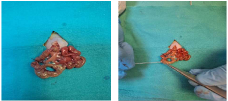
At 24 hrs after the experimental procedure, animals were sacrificed under ketamine (50 mg/kg) and xylazine (5 mg/kg) anesthesia by intracardiac blood collection, and heart and lung tissue samples were obtained (Fig. 1).
Histopathological analysis: Following fixation of the collected heart and lung tissue samples in 10% neutral formalin, routine tissue processing was performed, and paraffin blocks were prepared. Sections of 5 μm thickness obtained from the paraffin-embedded heart and lung tissues were stained with Hematoxylin and Eosin (H&E). The prepared slides were examined under a Leica DM 4000B (Germany) computer-assisted light microscope, and the obtained images were evaluated using the Leica LAS V4.9 software.
Based on light microscopic observations, histopathological changes in the heart and lung tissues were assessed semiquantitatively by scoring. In the histopathological evaluation of cardiac tissue, inflammation-neutrophil infiltration, myocardial disorganization, interstitial fibrosis, interstitial edema, myocardial cell swelling, and myocardial necrosis were examined. In the histopathological evaluation of lung tissue, interstitial edema, interstitial hemorrhage, alveolar congestion, neutrophil infiltration/aggregation, alveolar wall thickening, and total injury score were assessed.
Biochemical analysis: To evaluate oxidative stress and lipid peroxidation in cardiac and lung tissues, MDA levels and the activities of enzymes involved in free radical metabolism, including CAT enzyme activities, were assessed.
Statistical analysis: The study data were analyzed using the SPSS (Statistical Program for Social Sciences) software package. Statistical evaluation was performed with SPSS 20.0 using the tests listed below, and a p-value of <0.05 was considered statistically significant. Data are presented as Mean±Standard Deviation, median, and interquartile range (%25-%75). The Kolmogorov-Smirnov test was applied to measurable parameters to determine whether the distribution was normal or non-normal. Normally distributed data were expressed as Mean±Standard Deviation, while non-normally distributed data were presented as median and interquartile range (%25-%75). Differences between groups were analyzed using the Kruskal-Wallis test for independent groups. In cases where a significant difference was observed, pairwise comparisons were performed using the Mann-Whitney U test.
RESULTS
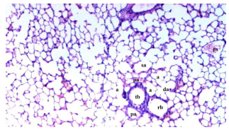
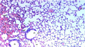
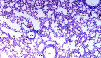
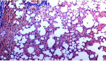
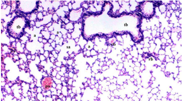
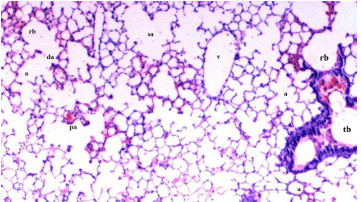
Interstitial edema was found to be significantly different among the groups (p = 0.026). Interstitial edema was observed more frequently in the DCLP group compared to the C, D, and DO groups (p = 0.001, p = 0.026, p = 0.007, respectively). Interstitial edema was significantly lower in the DCLPO and DOCLP groups compared to the DCLP group (p = 0.026, p = 0.007, respectively) (Table 1, Fig. 2-7).
Interstitial hemorrhage was significantly different among the groups (p = 0.007). Interstitial hemorrhage was more frequently observed in the DCLP group compared to the C, D, and DO groups (p<0.001, p = 0.042, p = 0.003, respectively). Moreover, interstitial hemorrhage was higher in the D and DCLPO groups compared to the C group (p = 0.042, both). Interstitial hemorrhage was significantly lower in the DCLPO and DOCLP groups compared to the DCLP group (p = 0.042, p = 0.012, respectively) (Table 1, Fig. 2-7).
Alveolar congestion was significantly different among the groups (p = 0.020). Alveolar congestion was observed more frequently in the DCLP group compared to the C and DO groups (p = 0.001, p = 0.007, respectively). Alveolar congestion was significantly lower in the DOCLP group compared to the DCLP group (p = 0.007) (Table 1, Fig. 2-7).
Neutrophil infiltration/aggregation was significantly different among the groups (p = 0.004). Neutrophil infiltration/aggregation was observed more frequently in the DCLP group compared to the C and DO groups (p<0.001, p = 0.010, respectively). In addition, neutrophil infiltration/aggregation was more frequent in the D and DCLPO groups compared to the C group (p = 0.002, p = 0.010, respectively). Neutrophil infiltration/aggregation was significantly lower in the DOCLP group compared to the DCLP group (p = 0.049) (Table 1, Fig. 2-7).
| Table 1: | Lung tissue histopathological findings (Mean±SE) | |||
| Group C (n = 6) |
Group D (n = 8) |
Group DO (n = 8) |
Group DCLP (n = 8) |
Group DCLPO (n = 8) |
Group DOCLP (n = 8) |
P** | |
| Interstitial edema | 0.50±0.22 | 0.83±0.17 | 0.67±0.21 | 1.50±0.22*,&,+ | 0.83±0.17? | 0.67±0.21? | 0.026 |
| Interstitial hemorrhage | 0.17±0.17 | 0.83±0.17* | 0.50±0.22 | 1.50±0.22*,&,+ | 0.83±0.17*,? | 0.67±0.33? | 0.007 |
| Alveolar congestion | 0.33±0.21 | 0.83±0.17 | 0.50±0.22 | 1.33±0.21*,+ | 0.83±0.17 | 0.50±0.22? | 0.02 |
| Neutrophil infiltration/aggregation | 0.17±0.17 | 1.00±0.00* | 0.50±0.22 | 1.17±0.17*,+ | 0.83±0.17* | 0.67±0.21? | 0.004 |
| Alveolar wall thickness | 0.33±0.21 | 1.17±0.17* | 0.33±0.21& | 1.83±0.40*,+ | 0.83±0.17? | 0.67±0.21? | <0.001 |
| Total damage score | 1.33±0.80 | 4.67±0.56* | 2.83±0.79 | 7.33±0.61*,&,+ | 4.17±0.40*,? | 3.17±0.54? | <0.001 |
| p**: Significance level with kruskal wallis test p<0.05 *p<0.05: Compared with group C, &p<0.05: Compared with group D, +p<0.05: Compared with Group DO, ?p<0.05: Compared with Group DCLP, C: Control group, D: Diabetes group, DO: Diabetes+Ozone group, DCLP: Diabetes+cecal ligation and perforation group, DCLPO: Diabetes+cecal ligation and perforation+ozone 1 group and DOCLP: Diabetes+ozone 2+cecal ligation and perforation group | |||||||
|
|
|
Alveolar wall thickness was significantly different among the groups (p<0.001). Alveolar wall thickness was observed more frequently in the DCLP group compared to the C and DO groups (p<0.001, both). Furthermore, alveolar wall thickness was greater in the D group compared to the C group (p = 0.021). Alveolar wall thickness was significantly lower in the DO group compared to the D group (p = 0.021). Similarly, alveolar wall thickness was significantly lower in the DCLPO and DOCLP groups compared to the DCLP group (p = 0.006, p = 0.002, respectively) (Table 1, Fig. 2-7).
The total injury score was significantly different among the groups (p<0.001). The total injury score was higher in the DCLP group compared to the C, D, and DO groups (p<0.001, p = 0.006, p<0.001,
|
|
| Table 2: | Heart tissue histopathological findings (Mean±SE) | |||
| Group C (n = 6) |
Group D (n = 8) |
Group DO (n = 8) |
Group DCLP (n = 8) |
Group DCLPO (n = 8) |
Group DOCLP (n = 8) |
P** | |
| Inflammation-neutrophil infiltration | 0.33±0.21 | 0.83±0.17 | 0.67±0.21 | 1.33±0.21*,+ | 0.83±0.17 | 0.67±0.21? | 0.036 |
| Myocardial disorganization | 0.33±0.21 | 0.83±0.17 | 0.67±0.21 | 1.17±0.17 | 0.83±0.31 | 0.67±0.21 | 0.194 |
| Interstitial fibrosis | 0.33±0.21 | 0.83±0.17 | 0.67±0.21 | 1.17±0.17 | 0.83±0.31 | 0.67±0.21 | 0.194 |
| Interstitial edema | 0.17±0.17 | 0.67±0.21 | 0.50±0.22 | 1.33±0.33*,&,+ | 0.83±0.17* | 0.67±0.21? | 0.029 |
| Swelling of myocardial cells | 0.33±0.21 | 1.00±0.26* | 0.50±0.22 | 1.33±0.21*,+ | 0.67±0.21? | 0.50±0.22? | 0.034 |
| Myocardial necrosis | 0.33±0.21 | 1.00±0.00* | 0.67±0.21 | 1.33±0.21*,+ | 0.67±0.21? | 0.67±0.21? | 0.02 |
| p**: Significance level with kruskal wallis test p<0.05 *p<0.05: Compared with group C, &p<0.05: Compared with group D, +p<0.05: Compared with group DO and ?p<0.05: Compared with Group DCLP | |||||||
respectively). In addition, the total injury score was higher in the D and DCLPO groups compared to the C group (p<0.001, p = 0.004, respectively). The total injury score was significantly lower in the DCLPO and DOCLP groups compared to the DCLP group (p = 0.001, p<0.001, respectively) (Table 1, Fig. 2-7).
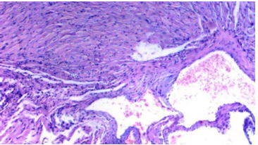
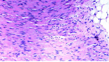
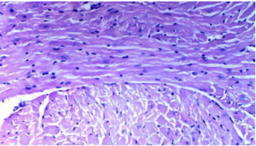
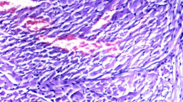
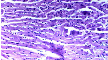
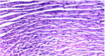
Inflammation neutrophil infiltration was found to be significantly different among the groups (p = 0.036). Inflammation neutrophil infiltration was observed more frequently in the DCLP group compared to the C and DO groups (p = 0.001, p = 0.023, respectively). Inflammation neutrophil infiltration was significantly lower in the DOCLP group compared to the DCLP group (p = 0.023) (Table 2, Fig. 8-13).
Interstitial edema was significantly different among the groups (p = 0.029). Interstitial edema was observed more frequently in the DCLP group compared to the C, D, and DO groups (p<0.001, p = 0.045, p = 0.014, respectively). In addition, interstitial edema was also higher in the DCLPO group compared to the C group (p = 0.045). Interstitial edema was significantly lower in the DOCLP group compared to the DCLP group (p = 0.045) (Table 2, Fig. 8-13).
|
|
Swelling in myocardial cells was significantly different among the groups (p = 0.034). Swelling in myocardial cells was observed more frequently in the DCLP group compared to the C and DO groups (p = 0.004, p = 0.013, respectively). Moreover, swelling in myocardial cells was also higher in the D group compared to the C group (p = 0.043). Swelling in myocardial cells was significantly lower in the DCLPO and DOCLP groups compared to the I/R group (p = 0.043, p = 0.013, respectively) (Table 2, Fig. 8-13).
Myocardial necrosis was significantly different among the groups (p = 0.020). Myocardial necrosis was more frequent in the I/R group compared to the C and O groups (p<0.001, p = 0.020, respectively). In addition, myocardial necrosis was higher in the D group compared to the C group (p = 0.020). Myocardial necrosis was significantly lower in the I/R+O and O+I/R groups compared to the I/R group (p = 0.020, both) (Table 2, Fig. 8-13).
Myocardial disorganization and interstitial fibrosis were found to be similar among the groups (p = 0.194, both) (Table 2, Fig. 8-13).
When the groups (lung tissue) were compared in terms of MDA levels, a significant difference was found among the groups (p = 0.002). The MDA level in the CLP group was significantly higher compared to the C, D, and DO groups (p<0.001, p = 0.013, p<0.001, respectively). In addition, the MDA level in the DCLPO group was significantly higher compared to the C group (p = 0.027). The MDA level in the DO group was significantly lower compared to the D group (p = 0.029). Similarly, the MDA levels in the DCLPO and DOCLP groups were significantly lower compared to the DCLP group (p = 0.024, p = 0.009, respectively) (Table 3).
|
|
|
| Table 3: | Lung tissue oxidant status parameters (Mean±SE) | |||
| Group C (n = 6) |
Group D (n = 8) |
Group DO (n = 8) |
Group DCLP (n = 8) |
Group DCLPO (n = 8) |
Group DOCLP (n = 8) |
P** | |
| MDA (nmol/g.pro) | 15.19±2.78 | 19.29±4.78 | 16.92±4.90 | 24.74±3.38*,&,+ | 20.40±4.97*,? | 17.82±3.51? | 0.002 |
| CAT (IU/mg.pro) | 52.85±3.12 | 51.21±9.49 | 54.13±5.21 | 38.96±5.98*,&,+ | 46.82±6.67+,? | 47.28±8.03? | 0.001 |
| p**: Significance level with kruskal wallis test p< 0.05 *p<0.05: Compared with group C, &p<0.05: Compared with group D, +p<0.05: Compared with group DO and ?p<0.05: Compared with group DCLP | |||||||
When the groups (lung tissue) were compared in terms of CAT enzyme activity, a significant difference was found among the groups (p<0.001). CAT enzyme activity in the CLP group was significantly lower compared to the C, D, and DO groups (p<0.001, all). In addition, CAT enzyme activity in the DCLPO group was significantly lower compared to the DO group (p = 0.039). CAT enzyme activity in the DCLPO and DOCLP groups was significantly higher compared to the DCLP group (p = 0.027, p = 0.020, respectively) (Table 3).
|
|
| Table 4: | Heart tissue oxidant status parameters (Mean±SE) | |||
| Group C (n = 6) |
Group D (n = 8) |
Group DO (n = 8) |
Group DCLP (n = 8) |
Group DCLPO (n = 8) |
Group DOCLP (n = 8) |
P** | |
| MDA (nmol/g.pro) | 62.78±6.85 | 87.70±13.13* | 74.31±6.50& | 106.00±17.47*,&,+ | 92.09±12.91*,+,? | 89.66±8.93*,+,? | <0.001 |
| CAT (IU/mg.pro) | 37.53±2.54 | 22.51±3.72* | 27.74±6.41*,& | 14.95±2.48*,&,+ | 20.97±4.38*,+,? | 21.34±3.33*,+,? | <0.001 |
| p**: Significance level with kruskal wallis test p<0.05 *p<0.05: Compared with group C, &p<0.05: Compared with group Dm, +p<0.05: Compared with group DO and ?p<0.05: Compared with group DCLP | |||||||
When the groups (heart tissue) were compared in terms of MDA levels, a significant difference was found among the groups (p<0.001). The MDA level in the DCLP group was significantly higher compared to the C, D, and DO groups (p<0.001, all). In addition, the MDA levels in the D, DCLPO, and DOCLP groups were also significantly higher compared to the C group (p<0.001, all). The MDA levels in the DCLPO and DOCLP groups were significantly lower compared to the DCLP group (p = 0.046, p = 0.002, respectively) (Table 4).
When the groups (heart tissue) were compared in terms of CAT enzyme activity, a significant difference was found among the groups (p<0.001). CAT enzyme activity in the DCLP group was significantly lower compared to the C, D, and DO groups (p<0.001, all). In addition, CAT enzyme activity in the D, DO, DCLPO, and DOCLP groups was also significantly lower compared to the C group (p<0.001, all). CAT enzyme activity in the DCLPO group was significantly higher compared to the DO and DCLP groups (p = 0.002, p = 0.005, respectively). Similarly, CAT enzyme activity in the DOCLP group was significantly higher compared to the DO and DCLP groups (p = 0.003, both) (Table 4).
DISCUSSION
Infection continues to be an important cause of morbidity and mortality in patients with diabetes and is most likely due to abnormalities in the host response, particularly in neutrophil chemotaxis and adhesion21. When reviewing the literature, we observe that although there are studies on the use of ozone in the treatment of sepsis, there are insufficient studies regarding its use in the treatment of sepsis developing in diabetic patients. In this study, we investigated the efficacy of ozone therapy against sepsis in diabetic subjects. We specifically evaluated the heart and lungs, which are the two organs most affected by sepsis.
In a study conducted by Park et al.22 in mice, it was shown that STZ-induced T1D was associated with the development of pulmonary fibrosis22. In our study, histological evaluation of lung tissue revealed that neutrophil infiltration/aggregation, alveolar wall thickness, and total injury score were higher in the diabetes group compared to the control group. This supports the notion that the lung is also one of the target organs of diabetes. In a study conducted by Park et al.22 on rats, gas exchange parameters were found to be impaired in both type 1 and type 2 DM models, with even greater deterioration in the type 1 DM model, indicating alveolo-capillary dysfunction23. In our study, the alveolar wall thickness, which was more pronounced in the diabetes group compared to the control group, was found to be lower in the ozone-treated DO group compared to the diabetes group. This suggests that alveolar wall thickening in the lungs caused by diabetes may be reversible with ozone therapy.
ALI results from the release of numerous inflammatory cytokines into the intracellular and extracellular matrix, leading to lung tissue injury, alveolar membrane damage, and alveolar-capillary diffusion dysfunction, ultimately causing persistent hypoxemia13,24. In a study conducted by Uzunçakmak et al.25 in a sepsis model (CLP group), they observed an increase in interalveolar septal thickening, desquamation of the bronchial epithelium, and venous hyperemia25. In the colon ascendens stent peritonitis (CASP) model established by Neumann and colleagues in mice, they found that CASP-induced abdominal sepsis leads to a massive accumulation of granulocytes in the lungs, and that this granulocyte accumulation is associated with a marked increase in pulmonary vascular permeability and increased expression of a group of chemokines and adhesion molecules26. In our study, the DCLP group showed higher levels of interstitial edema, interstitial hemorrhage, and total injury score compared with the diabetes group. This indicates that sepsis may also induce acute lung injury in diabetic mice.
In a study, it was shown that pantoprazole alleviated sepsis-induced acute lung injury and acute kidney injury in septic rats27. In another study, Fisher and colleagues demonstrated that parenteral vitamin C infusion in mice provided protection against harmful effects associated with sepsis, such as the proinflammatory response, impaired epithelial barrier function, alveolar fluid permeability, and sepsis-associated coagulopathy12. In a study on rats, de Souza et al.28 demonstrated that intraperitoneal ozone therapy, although not improving survival rates, potentially alleviated the inflammatory response and acute lung injury caused by intra-abdominal infection28. Similarly, in our study, we investigated whether ozone therapy could prevent sepsis-induced acute lung injury in diabetic mice. In lung histopathology, the DCLO group showed lower levels of interstitial edema, interstitial hemorrhage, alveolar wall thickening, and total injury score compared with the DCLP group. In the DOCLP group, interstitial edema, interstitial hemorrhage, alveolar congestion, neutrophil infiltration/aggregation, alveolar wall thickening, and total injury score were all lower compared with the DCLP group. From these findings, we conclude that ozone therapy may reduce sepsis-induced acute lung injury in diabetic mice. The observation that alveolar congestion and neutrophil infiltration/aggregation were reduced in the DOCLP group (ozone administered before CLP) but not in the DCLO group (ozone administered after CLP) suggests that preemptive ozone therapy is more effective in reducing acute lung injury.
In experimental studies, pathological changes in severe sepsis/septic shock such as disruption of the mechanical connection between the intracellular cytoskeleton and the extracellular matrix in cardiomyocytes, mechanical and electrical/chemical dissociation of cardiomyocytes, increased sarcolemmal permeability, and structural changes in the contractile apparatus of cardiomyocytes all of which have been associated with a decrease in myocardial contractile force29. In a study myocardial cells in the DM group were shown to be irregular, with an indistinct morphological structure and extensive inflammatory cell infiltration30. Another study demonstrated that all diabetic mice exhibited significant myocardial fibrosis in terms of biochemical and histopathological changes. In addition, structural and functional alterations at the myocardial level were also observed in diabetic mice. In that study, an 8-week empagliflozin treatment successfully preserved cardiac systolic function31. In our study, compared with the control group, the diabetes group exhibited significantly greater myocardial cell swelling and myocardial necrosis. However, no significant differences in myocardial disorganization or interstitial fibrosis were observed between the groups. Since we waited one month after inducing diabetes in mice to observe its effects, this period may not have been sufficient for the development of myocardial disorganization and interstitial fibrosis.
Patients with preexisting diabetes who develop sepsis experience a higher long-term risk of major cardiovascular events compared with non-diabetic sepsis patients. When compared with patients without sepsis, the absolute risk increase of cardiovascular events after sepsis is greater in diabetic patients32. Cai et al.29 induced sepsis with lipopolysaccharide (LPS) in STZ-induced diabetic mice. Histopathological examination of the heart revealed myocardial injury, including irregular arrangement, cardiomyocyte degeneration, and rupture of myocardial fibers. Furthermore, electron microscopy analysis demonstrated mitochondrial alterations in cardiomyocytes, vacuolization, and irregular swelling accompanied by partial myofibrillar dissolution33. In our study, greater interstitial edema was observed in the DCLP group compared with the diabetes group. When comparing the DCLP group with the control group, inflammation-neutrophil infiltration, myocardial cell swelling, interstitial edema, and myocardial necrosis were more pronounced in the DCLP group. These findings suggest that cardiac injury also occurs in the acute phase of sepsis in diabetic subjects, and if left untreated, this injury may lead to impaired myocardial contractility.
It is well known that sepsis is common in diabetic patients, and cardiac involvement in these patients increases morbidity, mortality, and length of stay in the intensive care unit. For this reason, numerous studies have been conducted to reduce sepsis-related cardiac involvement in diabetic patients. In a study by Cai et al.33 on diabetic mice, glibenclamide was shown to alleviate LPS-induced myocardial injury in STZ mice and to reduce serum IL-1β and TNF-α levels. They demonstrated that glibenclamide may provide protection against myocardial injury under inflammatory conditions in diabetes33. Ozone is known to have anti-inflammatory effects. We conducted this study to investigate whether ozone therapy could prevent inflammatory effects in cardiac tissue induced by sepsis. In our study, myocardial cell swelling and myocardial necrosis were found to be significantly lower in the DCLPO group compared with the DCLP group. Furthermore, in the DOCLP group, inflammation neutrophil infiltration, interstitial edema, myocardial cell swelling, and myocardial necrosis were significantly lower compared with the DCLP group. These findings suggest that ozone therapy may reduce inflammation in the heart. The observation that inflammation neutrophil infiltration and interstitial edema were lower in the DOCLP group but not in the DCLPO group indicates that preemptive administration of ozone is more effective in reducing cardiac inflammation.
In diabetic patients, in addition to increased capacity for ROS production, endogenous antioxidant mechanisms are frequently impaired34. In particular, ROS are produced within the mitochondria of the diabetic heart. The first line of defense against ROS-mediated cardiac injury consists of several antioxidant enzymes, including superoxide dismutase (SOD), CAT, and glutathione peroxidase (GSHPx)35. In our study, comparison of the control and diabetes groups revealed higher MDA levels and lower CAT enzyme activity in cardiac tissue, indicating that the effects of diabetes-related oxidative stress were more pronounced in the heart.
In animal models of diabetes, interventions with appropriate antioxidants such as SOD mimetics or coenzyme Q10 have demonstrated the causal role of oxidative stress in diabetic heart disease, either early as a preventive measure or later in the disease course, by limiting the progression to diabetic cardiomyopathy34. In a study by Khan et al.36 MDA levels were significantly reduced in the O3+Q10 group compared with the control group. Taken together, this study provided evidence of the beneficial effects of O3 therapy combined with Q10 in combating oxidation-induced injury and preventing such damage. Numerous studies have also provided evidence that O3 therapy enhances activation of the Nrf2 pathway through moderate induction of oxidative stress36. In our study, comparison of the control group with the DO group receiving ozone revealed significantly lower CAT enzyme activity in the heart, whereas MDA levels were similar between the groups. When comparing the diabetes and DO groups, a statistically significant reduction in MDA levels was observed only in lung tissue in the DO group. This suggests that ozone therapy is more effective on MDA, one of the oxidative stress markers associated with diabetes.
In a study conducted by Martinez-Sanchez et al.37 on patients with diabetic foot, improvements in glycemic control, a reduction in oxidative stress, an increase in SOD levels, significant healing of lesions, and a lower number of amputations compared to the control group were observed following ozone therapy. It has been demonstrated that O3 AHT improves blood circulation and oxygen delivery to ischemic tissues, reduces chronic oxidative stress by stimulating the antioxidant system, and may increase insulin secretion or efficacy37. In a study by Li et al.24 it was shown that in the DM group, MDA content increased while SOD activity significantly decreased; however, following simvastatin administration, SOD activity increased and MDA content was significantly reduced30. The results of these studies are consistent with and support our findings.
When the DM and CLP groups were compared, both lung and cardiac tissues showed high MDA levels and low CAT enzyme activity. These findings indicate that sepsis induces oxidative stress in both cardiac and pulmonary tissues. When the O3 and CLPO groups were compared, CAT enzyme activity in both lung and cardiac tissues was lower in the CLPO group, whereas no significant difference in MDA levels was observed between the two groups. When the O3 and DCLP groups were compared, CAT enzyme activity was found to be higher only in the cardiac tissue of the DCLP group. From these data, it can be concluded that ozone is more effective on CAT enzyme activity in the treatment of sepsis-induced oxidative stress.
In an experimental study conducted by Liu et al.24 on mice, they demonstrated that the CLP procedure contributes to lung injury by causing a significant decrease in serum SOD levels and an increase in MDA levels. Dexmedetomidine treatment significantly increased SOD levels and reduced MDA levels. Furthermore, it has been shown that dexmedetomidine alleviates ALI by increasing SOD enzyme activity while simultaneously reducing the release of inflammatory factors and MDA24. In another study conducted by Jacobs et al.38 on rats, it was demonstrated that increases in serum SOD and glutathione peroxidase (GSH-Px) levels in septic rats correlated well with improved survival38.
When the CLP and CLPO groups were compared, both cardiac and pulmonary tissues in the CLPO group showed lower MDA levels and higher CAT enzyme activity. When the CLP and DCLP groups were compared, both cardiac and pulmonary tissues in the DCLP group showed lower MDA levels and higher CAT enzyme activity. These findings demonstrate that ozone therapy can reduce oxidative stress caused by diabetes and sepsis in both cardiac and pulmonary tissues. Furthermore, in both groups where ozone was administered either before or after sepsis induction, a statistically significant reduction in oxidative stress was observed.
In the literature, a study conducted by Guanche et al.7 demonstrated that ozone oxidative preconditioning therapy at concentrations of 30-50 μg/mL protected the liver and lungs against oxidative stress and equally reduced tissue damage caused by experimental septic shock by maintaining the balance of pro-oxidant/antioxidant activities and reversing the effects of ROS7. Madej et al.39 using the well-known experimental model of endotoxemic shock induced by E. coli endotoxin in rats, also demonstrated the protective effects of ozone oxidative preconditioning in terms of SOD and CAT activities and their roles in controlling ROS levels39. In a study conducted by Yamanel et al.40 it was found that levels of the antioxidant enzymes SOD and GSH-Px were reduced in animals with sepsis; in the group that received ozone therapy, both SOD and GSH-Px activities were found to be significantly higher. Based on the results of this study, ozone therapy may serve as an alternative treatment approach to hyperbaric oxygen therapy in the treatment of sepsis, in addition to antibiotic therapy, due to its effects achieved through the modulation of the oxidative/antioxidative status40.
CONCLUSION
Histopathological and biochemical examinations of the tissues demonstrated that ozone therapy was able to reduce the effects of both diabetes and sepsis on the heart and lung tissues. We concluded that ozone therapy applied before the onset of sepsis was more effective than when applied after sepsis. We suggest that systemic ozone therapy could be considered as an adjuvant to antibiotic therapy in the treatment of sepsis in diabetic patients. Ozone therapy holds promise for reducing morbidity and mortality rates by mitigating the destructive effects of sepsis on the heart and lungs. We believe that further studies are required to elucidate the mechanisms, optimal dosage, and timing of ozone therapy to maximize its benefits in reversing sepsis-induced injury.
SIGNIFICANCE STATEMENT
This study demonstrates that ozone therapy may provide a protective adjunctive approach against cardiac and pulmonary tissue injury associated with sepsis in diabetes. The greater protective effect observed with pre-treatment suggests that the timing of ozone administration may be an important consideration in reducing oxidative stress and inflammation during sepsis. These findings provide experimental evidence supporting further investigation of ozone therapy as a potential adjunctive strategy for managing sepsis-related organ damage in diabetic conditions.
REFERENCES
- Loscalzo, J., A.S. Fauci, D.L. Kasper, S. Hauser, D. Longo and J.L. Jameson, 2022. Harrison's Principles of Internal Medicine. 21st Edn., McGraw Hill LLC, New York, USA, ISBN: 9781264268511, Pages: 4384.
- Bodke, H., V. Wagh and G. Kakar, 2023. Diabetes mellitus and prevalence of other comorbid conditions: A systematic review. Cureus, 15.
- Trevelin, S.C., D. Carlos, M. Beretta, J.S. da Silva and F.Q. Cunha, 2017. Diabetes mellitus and sepsis: A challenging association. Shock, 47: 276-287.
- Şengel, N., A. Küçük, Ç. Özdemir, Ş.C. Sezen and G. Kip et al., 2023. The effect of sevoflurane and fullerenol C 60 on the liver and kidney in lower extremity ischemia-reperfusion injury in mice with streptozocin-induced diabetes. Int. J. Nanomed., 18: 7543-7557.
- Marino, P.L., 2024. Marino's The ICU Book: EBook Without Updates. Wolters Kluwer Health, Philadelphia, Pennsylvania, United States, ISBN: 9781975176716 Pages: 864.
- Singer, M., C.S. Deutschman, C.W. Seymour, M. Shankar-Hari and D. Annane et al., 2016. The third international consensus definitions for sepsis and septic shock (Sepsis-3). J. Am. Med. Assoc., 315: 801-810.
- Guanche, D., F. Hernandez, Z. Zamora and Y. Alonso, 2010. Effect of ozone pre-conditioning on redox activity in a rat model of septic shock. Toxicol. Mech. Methods, 20: 466-471.
- Skorup, P., A. Fransson, J. Gustavsson, J. Sjöholm and H. Rundgren et al., 2022. Evaluation of an extracorporeal ozone-based bactericide system for the treatment of Escherichia coli sepsis. Intensive Care Med. Exp., 10.
- Lilly, C.M., R.S. Irwin, W.A. Boyle and W.F. Kelly, 2023. Irwin and Rippe's Intensive Care Medicine: EBook Without Multimedia. Wolters Kluwer Health, Philadelphia, Pennsylvania, United States, ISBN: 9781975181468, Pages: 2,400.
- Iskander, K.N., F.L. Craciun, D.M. Stepien, E.R. Duffy and J. Kim et al., 2013. Cecal ligation and puncture-induced murine sepsis does not cause lung injury. Crit. Care Med., 41: 159-170.
- Filgueiras Jr, L.R., J.O. Martins, C.H. Serezani, V.L. Capelozzi, M.B.A. Montes and S. Jancar, 2012. Sepsis-induced acute lung injury (ALI) is milder in diabetic rats and correlates with impaired NFkB activation. PLoS ONE, 7.
- Fisher, B.J., D. Kraskauskas, E.J. Martin, D. Farkas and J.A. Wegelin et al., 2012. Mechanisms of attenuation of abdominal sepsis induced acute lung injury by ascorbic acid. Am. J. Physiol. Lung Cell. Mol. Physiol., 303: L20-L32.
- Jin, L.Y., C.F. Li, G.F. Zhu, C.T. Wu, J. Wang and S.F. Yan, 2014. Effect of siRNA against NF-κB on sepsis-induced acute lung injury in a mouse model. Mol. Med. Rep., 10: 631-637.
- Sorgun, O., A. Çakır, E.S. Bora, M.A. Erdoğan, Y. Uyanıkgil and O. Erbaş, 2023. Anti-inflammatory and antioxidant properties of betaine protect against sepsis-induced acute lung injury: CT and histological evidence. Braz. J. Med. Biol. Res., 56.
- Frijhoff, J., P.G. Winyard, N. Zarkovic, S.S. Davies and R. Stocker et al., 2015. Clinical relevance of biomarkers of oxidative stress. Antioxid. Redox Signaling, 23: 1144-1170.
- Halliwell, B., 2006. Reactive species and antioxidants. Redox biology is a fundamental theme of aerobic life. Plant Physiol., 141: 312-322.
- Orakdogen, M., S. Uslu, S.T. Emon, H. Somay, Z.C. Meric and T. Hakan, 2016. The effect of ozone therapy on experimental vasospasm in the rat femoral artery. Turk. Neurosurg., 26: 860-865.
- Gülcan, M.B., H. Demirtaş, A. Özer, Z. Yığman, A.D. Dursun, M. Arslan and G.L. Oktar, 2024. Ozone administration reduces myocardial ischemia reperfusion injury in streptozotocin induced diabetes mellitus rat model. Drug Des. Dev. Ther., 18: 4203-4213.
- Liu, L., L. Zeng, L. Gao, J. Zeng and J. Lu, 2023. Ozone therapy for skin diseases: Cellular and molecular mechanisms. Int. Wound J., 20: 2376-2385.
- de Sire, A., F. Agostini, L. Lippi, M. Mangone and S. Marchese et al., 2021. Oxygen-ozone therapy in the rehabilitation field: State of the art on mechanisms of action, safety and effectiveness in patients with musculoskeletal disorders. Biomolecules, 11.
- Koh, G.C.K.W., S.J. Peacock, T. van der Poll and W.J. Wiersinga, 2012. The impact of diabetes on the pathogenesis of sepsis. Eur. J. Clin. Microbiol. Infect. Dis., 31: 379-388.
- Park, S.S., M. Mai, M. Ploszaj, H. Cai and L. McGarvey et al., 2023. Type 1 diabetes contributes to combined pulmonary fibrosis and emphysema in male alpha 1 antitrypsin deficient mice. PLoS ONE, 18.
- Südy, R., Á. Schranc, G.H. Fodor, J. Tolnai, B. Babik and F. Peták, 2020. Lung volume dependence of respiratory function in rodent models of diabetes mellitus. Respir. Res., 21.
- Liu, Z., J. Xu, Y. Zhao, Y. Wan and R. Guo et al., 2024. Dexmedetomidine protects against sepsis-induced lung injury through autophagy and Smad2/3 signaling pathway. Iran. J. Basic Med. Sci., 27: 456-460.
- Uzunçakmak, S.K., Z. Halıcı, S. Karakaya, Z. Kutlu and Y.S. Sağlam et al., 2023. Suberosin alleviates sepsis-induced lung injury in a rat model of cecal ligation and puncture. J. Invest. Surg., 36.
- Neumann, B., N. Zantl, A. Veihelmann, K. Emmanuilidis, K. Pfeffer, C.D. Heidecke and B. Holzmann, 1999. Mechanisms of acute inflammatory lung injury induced by abdominal sepsis. Int. Immunol., 11: 217-227.
- Cao, L., F. Zou, C. Zhang, K. Xu, J. Zhao and J. Liu, 2027. CREG1 mitigates sepsis-induced acute lung injury by mediating macrophage polarization via AMPK/NF-κB signaling. Tissue Cell, 104.
- de Souza, Y.M., B. Fontes, J.O. Martins, P. Sannomiya, G.S. Brito, R.N. Younes and S. Rasslan, 2010. Evaluation of the effects of ozone therapy in the treatment of intra-abdominal infection in rats. Clinics, 65: 195-202.
- Celes, M.R.N., C.M. Prado and M.A. Rossi, 2013. Sepsis: going to the heart of the matter. Pathobiology, 80: 70-86.
- Al-Rasheed, N.M., N.M. Al-Rasheed, I.H. Hasan, M.A. Al-Amin, H.N. Al-Ajmi, R.A. Mohamad and A.M. Mahmoud, 2017. Simvastatin ameliorates diabetic cardiomyopathy by attenuating oxidative stress and inflammation in rats. Oxid. Med. Cell. Longevity, 2017.
- Li, C., J. Zhang, M. Xue, X. Li and F. Han et al., 2019. SGLT2 inhibition with empagliflozin attenuates myocardial oxidative stress and fibrosis in diabetic mice heart. Cardiovasc. Diabetology, 18.
- Angriman, F., P.R. Lawler, B.R. Shah, C.M. Martin and D.C. Scales, 2023. Prevalent diabetes and long-term cardiovascular outcomes in adult sepsis survivors: A population-based cohort study. Crit. Care, 27.
- Cai, J., S. Lu, Z. Yao, Y.P. Deng and L.D. Zhang et al., 2013. Glibenclamide attenuates myocardial injury by lipopolysaccharides in streptozotocin-induced diabetic mice. Cardiovasc. Diabetol., 14.
- Ritchie, R.H. and E.D. Abel, 2020. Basic mechanisms of diabetic heart disease. Circ. Res., 126: 1501-1525.
- Tsutsui, H., S. Kinugawa, S. Matsushima and T. Yokota, 2010. Oxidative stress in cardiac and skeletal muscle dysfunction associated with diabetes mellitus. J. Clin. Biochem. Nutr. 48: 68-71.
- Khan, S.A., N.L. Smith, A.L. Wilson, J. Gandhi and S. Vatsia, 2017. Ozone therapy: An overview of pharmacodynamics, current research, and clinical utility. Med. Gas Res., 7: 212-219.
- Martinez-Sanchez, G., S.M. Al-Dalain and S. Menendez, 2005. Therapeutic efficacy of ozone in patients with diabetic foot. Eur. J. Pharmacol., 523: 151-161.
- Jacobs, S., S. Sobki, C. Morais and M. Tariq, 2000. Effect of pentaglobin and piperacillin on survival in a rat model of faecal peritonitis: Importance of intervention timings. Acta Anaesthesiologica Scand., 44: 88-95.
- Madej, P., A. Plewka, J.A. Madej, M. Nowak, D. Plewka, G. Franik and D. Golka, 2007. Ozonotherapy in an induced septic shock. I. Effect of ozonotherapy on rat organs in evaluation of free radical reactions and selected enzymatic systems. Inflammation, 30: 52-58.
- Yamanel, L., U. Kaldirim, Y. Oztas, O. Coskun and Y. Poyrazoglu et al., 2011. Ozone therapy and hyperbaric oxygen treatment in lung injury in septic rats. Int. J. Med. Sci., 8: 48-55.
How to Cite this paper?
APA-7 Style
Şimşek, B. H., Göbüt, H., İnan, N., Arslan, M., Dikmen, K., Sezen, Ş. C., Kavutçu, M., & Küçük, A. (2026). Effects of ozone therapy on cardiac and pulmonary tissues in cecal perforation-induced sepsis in streptozotocin-induced diabetic mice. Asian Journal of Biological Sciences, 19(4), 134–149. https://doi.org/10.3923/ajbs.2026.134.149
ACS Style
Şimşek, B. H.; Göbüt, H.; İnan, N.; Arslan, M.; Dikmen, K.; Sezen, Ş. C.; Kavutçu, M.; Küçük, A. Effects of Ozone Therapy on Cardiac and Pulmonary Tissues in Cecal Perforation-Induced Sepsis in Streptozotocin-Induced Diabetic Mice. Asian J. Biol. Sci 2026, 19 (4), 134–149. https://doi.org/10.3923/ajbs.2026.134.149
AMA Style
Şimşek BH, Göbüt H, İnan N, et al. Effects of ozone therapy on cardiac and pulmonary tissues in cecal perforation-induced sepsis in streptozotocin-induced diabetic mice. Asian Journal of Biological Sciences. 2026;19(4):134–149. doi:10.3923/ajbs.2026.134.149
Chicago/Turabian Style
Şimşek, Bilgin Hasret, Hüseyin Göbüt, Nurten İnan, et al. 2026. “Effects of Ozone Therapy on Cardiac and Pulmonary Tissues in Cecal Perforation-Induced Sepsis in Streptozotocin-Induced Diabetic Mice.” Asian Journal of Biological Sciences 19 (4): 134–149. https://doi.org/10.3923/ajbs.2026.134.149

This work is licensed under a Creative Commons Attribution 4.0 International License.

