Cocos nucifera Water Ameliorated Hepatic Complications and Attenuated Oxidative Stress in Cadmium-Induced Hepatotoxicity
-
Solomon Kingsley Nwadum
Department of Pharmacology and Therapeutics, Faculty of Clinical Basic Medicine, Ebonyi State University, Abakaliki, Ebonyi, Nigeria
Udu Ama IbiamDepartment of Biochemistry, Faculty of Science, Ebonyi State University, Abakaliki, Ebonyi, Nigeria
Daniel Ejim Uti

Department of Biochemistry, Faculty of Basic Medical Sciences, College of Medicine, Federal University of Health Sciences Otukpo, Otukpo, Benue, Nigeria
Grace Ufedo UmoruDepartment of Biochemistry, College of Science, Evangel University Akaeze, Akaeze, Ebonyi, Nigeria
Mfon Paulinus UdoudohDepartment of Basic Sciences, Federal College of Medical Laboratory Science and Technology, Jos, Plateau, Nigeria
Patrick Maduabuchi AjaDepartment of Biochemistry, Faculty of Science, Ebonyi State University, Abakaliki, Ebonyi, Nigeria
Esther Ugo AlumDepartment of Biochemistry, Faculty of Science, Ebonyi State University, Abakaliki, Ebonyi, Nigeria
Chukwufumnanya Joseph MordiDepartment of Medical Biochemistry, Delta State University, Abraka, Delta, Nigeria
Ezebuilo Ugbala EkponoDepartment of Biochemistry, Federal Polytechnic, Oko, Anambra, Nigeria
Uket Nta ObetenDepartment of Chemistry, Biochemistry and Molecular Biology, Alex Ekwueme Federal University, Abakaliki, Ebonyi, Nigeria
Wilson Achu OmangDepartment of Medical Laboratory Science, Cross River State College of Health Technology, Calabar, Cross River State, Nigeria
Samuel Ali AgadaDepartment of Biochemistry, Faculty of Basic Medical Sciences, College of Medicine, Federal University of Health Sciences Otukpo, Otukpo, Benue, Nigeria
| Received 15 Sep, 2023 |
Accepted 19 Nov, 2023 |
Published 31 Dec, 2023 |
Background and Objectives: Cadmium (Cd) is a heavy metal widely distributed in the environment due to industrial, agricultural and natural sources. Chronic exposure to cadmium is known to induce hepatotoxicity, leading to severe liver damage and dysfunction. Hence, the effect of Cocos nucifera water on indices of liver function and oxidative stress in cadmium-induced hepatotoxicity was investigated in albino rats. Materials and Methods: The study involved 30 Wistar rats divided into 6 groups, with control groups receiving normal saline. Hepatotoxicity was induced in groups B, D, E and F by oral cadmium administration. Group B remained untreated, while groups D through F were treated with varying doses of Cocos nuciferawater. The proximate, vitamin and mineral contents of Cocos nuciferawater were determined. Results: The cadmium administration to rats increased hepatic enzyme activities, decreased catalase and reduced glutathione levels. However, treatment with Cocos nucifera water significantly decreased these effects and restored liver architecture to normal. The study confirmed that cadmium can be a potential health risk for humans. Conclusion: The Cocos nucifera water maybe beneficial in treating liver complications.
| Copyright © 2023 Nwadum et al. This is an open-access article distributed under the Creative Commons Attribution License, which permits unrestricted use, distribution, and reproduction in any medium, provided the original work is properly cited. |
INTRODUCTION
Plant resources have remained an integral part of human society throughout history. After having met the primary needs such as food and shelter, men searched for a suitable remedy among the plants to cure various diseases1.
Cocos nucifera palms are typically tall, unbranched and monoecious trees. They have a smooth, columnar trunk that is light grey-brown in color and can reach heights of 9-18 m or even up to 30 m in some cases. There are also dwarf varieties of these palms. The coconut fruit is composed of several layers starting from the outermost: A thin, hard shell known as the exocarp, followed by a thicker layer of fibrous mesocarp referred to as the shell. Beneath these layers, there is the hard endocarp, often called the shell as well and then the white endosperm, which is the kernel. Within the coconut, there is also a sizable cavity that contains a watery fluid, commonly known as coconut water or milk. When the endosperm is immature, it is soft and gelatinous but it becomes firm as it matures. In unripe coconuts, there is an abundance of coconut water or milk, which gradually gets absorbed as the coconut ripens. The color of the fruits changes from green when they are young to brownish as they ripen, while yellow varieties transition from yellow to brown as they mature.
Cocos nucifera water consists of 95.5% water, 4% sugar, 0.1% fat, 0.02% calcium, 0.01% phosphorus, 0.5% iron, considerable amounts of amino acids, mineral salts, vitamin B complex, vitamin C and cytokines etc.2,3. Other components in Cocos nucifera water are sugar (glucose), sugar alcohols, lipids, amino acids, nitrogen compounds, organic acids and enzymes4. The characteristic aroma of Cocos nucifera is contributed by deltalactones5. The nutritional composition of Cocos nucifera water is however, influenced by several factors, including the state of maturity, soil and environmental conditions.
Cadmium (Cd), a hazardous and non-essential element, finds extensive application in electroplating, pigments, paints, welding and batteries, impacting both living and non-living environments. In stark contrast to organic compounds, Cd does not naturally break down and possesses an extraordinarily long biological half-life. Numerous studies have established that Cd triggers a broad spectrum of biochemical and physiological disruptions in humans, laboratory animals and plants6-8. Various organs in mammals, including the kidney, liver, testes, lungs, pancreas, prostate, ovaries and placenta, are vulnerable to Cd’s detrimental effects. Valko et al.9 highlighted Cd’s adverse impact on spermatogenesis, primarily through the generation of free radicals. Substantial evidence also indicates that Cd disrupts antioxidant defense systems and elevates the production of reactive oxygen species (ROS) within cells, encompassing singlet oxygen, hydrogen peroxide and hydroxyl radicals9. When these ROS interact with macromolecules, they induce oxidative stress within cells, leading to a range of damage, such as DNA mutations, disruption of protein function and structure, lipid peroxidation, alterations in gene expression and apoptosis10. Monitoring tissue levels of malondialdehyde (MDA) and the activities of superoxide dismutase (SOD), glutathione peroxidase (GPx) and catalase (CAT) serves as established indicators of oxidative stress11. Chang et al.12 delved into the impact of Cd on the alteration of antioxidative defense systems and the induction of apoptosis in the hepato-pancreas of S. henanense, while Wang et al.13 proposed a potential link between Cd-induced apoptosis and oxidative stress. Numerous reports have consistently underscored the pivotal role of oxidative stress as a key mechanism underlying Cd’s toxicity14.
Cocos nucifera water has several culinary uses. It is renowned in Traditional Chinese Medicine for bolstering the heart and boosting endurance and its oils are used as insects repellants in Chinese culture3,15.
Numerous research studies have demonstrated that Cocos nucifera water possesses antiviral, antibacterial, anti-inflammatory and antioxidant attributes, which can be beneficial in alleviating a wide spectrum of mild to severe health issues. This nutrient-rich beverage has been employed to help regulate blood pressure, blood sugar and cholesterol levels, as well as to enhance energy levels and boost metabolism in the human body3. Furthermore, it has shown promise in addressing various ailments such as stomach flu, dysentery, indigestion, constipation, intestinal worms, urethral stones, kidney dysfunction, dry and itchy skin, age spots and wrinkles16. Cocos nucifera water can also elevate high-density lipoprotein (HDL) cholesterol, making it a natural remedy for maintaining cardiovascular health. Notably, young Cocos nucifera water exhibits estrogen-like properties and was even used for emergency transfusions during World War II due to its compatibility with blood17. Additionally, Cocos nucifera water can serve as a short-term intravenous fluid source, thanks to its high sugar and salt content, similar to modern intravenous solutions like lactate Ringer’s or dextrose water solutions18.
In Eastern Nigeria, Cocos nucifera water is used for various medicinal purposes, including the treatment and management of various conditions such as gastrointestinal disorders, high blood pressure, dehydration, kidney dysfunction, anxiety, etc.2. Hence, the motivation behind this study on Cocos nucifera water in cadmium induced hepatotoxic rats was to evaluate the hepatoprotective effects of Cocos nucifera water in a cadmium-induced hepatotoxicity model and to assess the impact of Cocos nucifera water on oxidative stress markers in the liver.
MATERIALS AND METHODS
The initial steps involved in preparing the nuts and conducting the analysis took 3 weeks. The phase focused on exposing the subjects to cadmium spanned 2 weeks and the treatment with Cocos nucifera water (administered once daily) continued for 28 days, equivalent to 4 weeks. Altogether, these activities extended the study duration to a total of 9 weeks before data collection and analysis from the animal experiment. These processes took place at the Department of Biochemistry, Ebonyi State University, Abakaliki, Ebonyi State, Nigeria from August to October, 2022.
Chemicals and reagents: Chemicals used in this study were of high analytical grade and were bought from Sigma Aldrich (St. Louis, Morocco, United States of America) and the reagents used for some assays were commercial kits and products of Randox Laboratories Ltd., United Kingdom.
Collection of nuts: In Mgbabor in Abakaliki, Ebonyi State, Nigeria, a total of 30 Cocos nucifera nuts (all same type, green high) were collected. The Cocos nucifera nuts were identified and authenticated by a taxonomist from the Department of Applied Biology at Ebonyi State University, Abakaliki. The Cocos nucifera was carefully collected by draining it through drilled holes through the mycrophylls and the water was collected in the beaker and used immediately. The water was carefully harvested and checked to ensure that there were no peel residues in it and given fresh19.
Proximate, mineral and vitamin analysis of Cocos nucifera water: Percentage compositions of protein, ash, moisture and carbohydrates were determined using the method of the Association of Official Analytical Chemists A.O.A.C.20, while the fat content was determined by the gravimetric solvent extraction method described by James21. Moreover, mineral composition, potassium, calcium, magnesium and iron were performed by the method of Moyo et al.22 while the vitamins were determined according to the methods of A.O.A.C.20.
Experimental animals and design
Experimental animals: A total of thirty albino rats (100-190 g) were obtained from Animal House, University of Nigeria, Nsukka, Enugu State. They were housed under standard laboratory conditions for 2 weeks for acclimatization and fed rodent chow and water ad libitum in the Animal House of Biochemistry Department at Ebonyi State University in Abakaliki. The animals were divided into 6 groups (n = 5). Group A served as a normal control given ad libitum with rat chow and water. Toxicity was induced in groups B, D, E and F by oral administration of 15 mg kg– b.wt., cadmium for 14 consecutive days. Group B, which served as a positive control, was left without treatment. Group C received 2.0 mL kg–1 of Cocos nucifera water via oral gavage without induction. Groups D, E and F were treated with 1.0, 2.0 and 3.0 mL kg–1 of Cocos nucifera water, respectively following an earlier report by Agbafor et al.23 with minor modifications. The cadmium exposure (14 days) and treatment with Cocos nucifera water (once per day) lasted for 28 days (4 weeks). The weight of the rats was measured and recorded before and after the experimental treatments. Animals were sacrificed and blood samples drawn from the animals after an overnight fast by cardiac puncture under light anesthesia using diethyl ether.
Determination of liver function parameters: The activities of Alanine Aminotransferase (ALT) and Aspartate Aminotransferase (AST) were assayed according to the method of Reitman and Frankel24, while Alkaline Phosphatase (ALP) activity was assayed by colorimetric method as described by Meulemans25.
Determination of oxidative stress parameters
Determination of malondialdehyde (MDA) level: The MDA was determined by spectrophotometric method as described by Wallin et al.26. In a nutshell, the test tube was filled with exactly 0.1 mL of sample, 0.9 mL of distilled water, 0.5 mL of TCA reagent and 0.5 mL of TBA reagent before being sealed. The test tube was incubated for 40 min at 95°C. After that, the test tube was allowed to cool in water for a few minutes before a precise 0.1 mL of SDS (sodium dodecyl sulphate) solution was added to it. The absorbance of the sample was measured at 532 and 600 nm in comparison to the blank reagent.
Calculation:
 |
Determination of catalase (CAT) activity: The CAT activity was assayed according to the method described by Udeozor et al.27. In brief, 2.5 mL of phosphate buffer and 2 mL of H2O2 were added to the test tube exactly at the same times. Following that, 0.5 mL of the sample was also put to the test tube. The 2 mL of dichromate acetic acid reagent were added to a 1 mL aliquot of the reaction mixture. The absorbance was measured at 240 nm against a blank at a rate of 1 reading per min:
 |
Where:
| A | = | Change in absorbance | |
| VT | = | Total volume | |
| Vs | = | Sample volume | |
| Σ | = | Molar extinction |
Determination of the SOD activity: The method described by Zima and Kalousová28 was used to determine the activity of SOD. Based on the inhibition of nitrobuletetrazolium (NBT) reduction. The sample was mixed with 2.95 mL of 0.05 M sodium carbonate buffer (pH 10.2) and 0.03 mL of epinephrine in 0.005 M HCl to begin the reaction, which took 0.02 mL in total. Similarly, the blank had 2.95 mL buffer, 0.03 mL of epinephrine and 0.02 mL of distilled water. The SOD activity was determined by measuring the change in absorbance at 480 nm for 5 min with a spectrophotometer. '= 4020 M–1 cm–1.
Calculation:
 |
Where:
| ΔA | = | Change in absorbance | |
| VT | = | Total volume | |
| Vs | = | Sample volume | |
| Σ | = | Molar extinction |
Determination of reduced GSH: This was determined using the method of Ellman29. Reduced GSH constitutes the vast majority of non-protein sulfhydryl groups in this assay. The procedure was straightforward: 0.2 mL of serum was combined with 2 mL of 10% trichloroacetic acid and centrifuged to separate the protein. Then 2 mL of phosphate buffer (pH 8.4), 0.5 mL of 5, 5-dithio, bis (2-nitrobenzoic acid) and 0.4 mL of double distilled water to 0.01 mL of the supernatant to achieve the desired pH. The absorbance was read at 412 nm after the mixture had been vortexed. The concentration of GSH was calculated as follows:
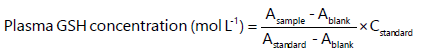 |
Histological examination of hepatic tissue: For brevity, in this investigation, liver tissue was fixed in 10% formal saline for three days in a container with light-tight lids to prevent autolysis, increase staining quality and help optical differentiation of cells. The tissue was dried to remove water that was not miscible with xylene and wax using different grades of alcohol ranging from 50% to absolute alcohol for 30 min on each side for a total of 3 hrs. After the dehydrated tissue had been cleaned, it was immersed in three changes of xylene for 30 min each to remove the alcohol that had accumulated on it. A hot oven (high temperature laboratory oven range LHT) and a temperature of 55°C was used to impregnate and penetrate the cleared tissue to remove the clearing agent (xylene). The tissue was passed through three changes of molten paraffin wax in a hot air oven for 30 min to remove the clearing agent. It was necessary to bury or embed the infiltrating tissue in molten paraffin wax in an embedded mold before the wax could be solidified. It was necessary to adhere a paraffin block of tissue to a wooding block using a hot spatula that was sandwiched between the wooding block and the paraffin wax. It is necessary to remove the spatula for the wax to be melted and solidified. To stain the tissue, it was sectioned using a rotary microtome (Manual Rotatory Microtome 0.5-60 μm (Code: 690.011) with precise advancement system, cutting range from 0.5 to 60 μm) and trimmed to 15 microns to acquire the cutting surface of the tissue. The tissue was then sectioned at 5 microns and dried in a hot plate before staining was completed. The segment was hydrated and then cleared with xylene to complete the process. After that, it was immersed in hematoxylin. After being cleaned with tap water, the portion was immersed in an acid-alcohol solution for 15 min. The piece was soaked in eosin stain for 15 min, after which the excess stain was washed away with tap water. The section was dehydrated with ethanol and mounted on a microscope slide using a resinous medium containing emission oil, after which the cells were examined.
Statistical analysis: The data obtained were expressed as Mean±STD and subjected to One-Way Analysis of Variance (ANOVA) followed by Duncan’s Multiple Range comparison and Prism GraphPad 7. Values p<0.05 were considered significant.
RESULTS
Chemical composition of Cocos nucifera water: The results of the proximate composition of Cocos nucifera water showed the following order of occurrence: Moisture>carbohydrates>ash>fat>protein (Fig. 1). Potassium content was observed to have the highest value followed by magnesium, calcium and the least was iron in mineral composition (Fig. 2). The vitamin content of Cocos nucifera water revealed the following order of occurrence vitamin C>B9>E>B1, while vitamin A was completely absent (Fig. 3).

|

|

|

|
|
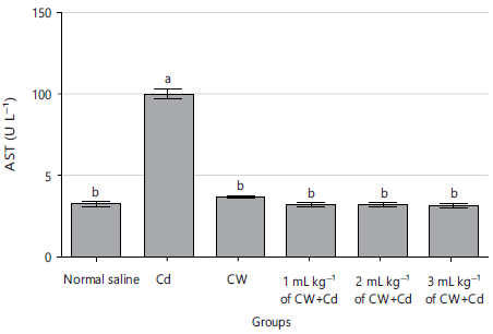
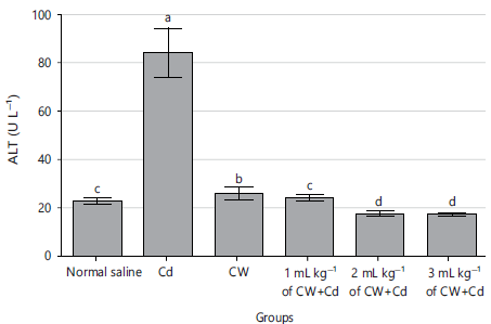
Effect of Cocos nucifera water on liver enzymes of Cd-induced hepato-toxicity in albino rats: The results on liver function enzymes in Cd-induced hepatotoxicity rats showed a significant (p<0.05) increase in ALP, AST and ALT levels in the groups that received 15 mg kg–1 (b.wt.) of Cd only relative to other groups as shown in Fig. 4-6. However, the administration of rats with different doses of Cocos nucifera water caused a reversal of the trend significantly (p<0.05) reducing the activity of the liver enzyme parameters to levels similar to the normal control (Fig. 4-6).
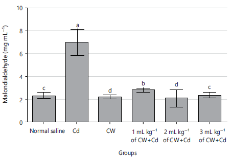
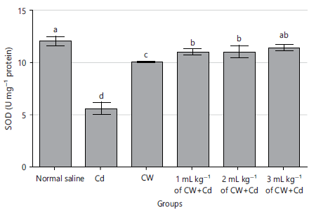
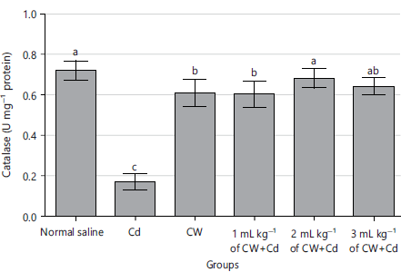
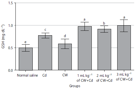
Effect of Cocos nucifera water on oxidative stress indices in Cd-induced hepato-toxicity in albino rats: The results on oxidative stress index in Cd-induced hepato-toxicity in albino rats showed a significant (p<0.05) increase in MDA level (Fig. 7) and a significant (p<0.05) decrease in the levels of SOD, catalase and GSH indices as shown in Fig. 8-10 on administration of 15 mg kg–1 (b.wt.) of Cd. However, treatment of the rats with different doses of Cocos nucifera water caused a reversal of the trend significantly (p<0.05) reducing the activity of the oxidative parameters to levels like the normal control. However, the effect was not dose-dependent for SOD activity.
|
|
Effect of Cocos nucifera water on histopathology of liver in Cd induced hepato-toxicity in albino rats: The results on histopathological effect on the liver showed severe aggregate of inflammatory cells (AIC) around the hemorrhagic area, indicating severe chronic hepatitis features as shown in Fig. 11. The liver cells showed well perfused normal lobular architecture with central vaen (CV), portal triad (PT) and hepatocytes (H) with mild aggregate of inflammatory cells (AIC) within the portal traid otherwise normal as shown in Plates 1 in Fig. 11. Plate 2 on administration of 15 mg kg–1 b.wt., of Cd. However, treatment of cadmium intoxicated albino rats with different doses of Cocos nucifera water caused a moderately healing effect with mild aggregates of intra lobular inflammation (AILI) around the intra hepatic hemorrhage (IHH) in r1 and mild congestion of the portal vain (CPV) as shown in Plates 3-6.
|
|
|

|
DISCUSSION
This study investigated the impact of Cocos nucifera water in cadmium-induced hepatotoxicity. The proximate, mineral and vitamin contents of C. nucifera water were quite fascinating. Moisture, carbs, ash, fat and protein were all detected in considerable amounts. This was in agreement with an earlier report by Kannaian et al.30 showing that C. nucifera water contains crude protein, crude fat, moisture, ash and carbohydrates. Nonetheless, the current study found some changes in the amounts of various proximate components, which could be due to differences in soil factors at different geographical sites where plants were obtained for proximate research. Even though lipids and proteins were also present in substantial proportions, our findings demonstrated that C. nucifera water is a better source of carbohydrates than the three basic macromolecules investigated (carbohydrates, lipids and protein). The high quantities of lipids and carbohydrates in C. nucifera water may explain its widespread use in nutrition around the world, as well as its utility as a source of energy for a variety of biological processes. Because of the high protein content of C. nucifera water, it may have important immunological, physiological, nutritional and pharmacological impacts on the body, contributing to general health and well-being. The relatively high composition of protein, carbohydrate and fats present in C. nucifera water indicates that the plant could be beneficial in both human and livestock nutrition31.
Mineral content indicated potassium to have the highest value followed by magnesium, calcium and iron. The finding of this study was similar to work of Imo et al.32, who reported the amount of potassium, magnesium and sodium to be higher than other minerals evaluated in C. nucifera water. This work also relates closely to the report of Evuen et al.33, who estimated the nutritive potentials of C. nucifera water as magnesium, calcium, potassium and iron were all present. However, the mineral contents of C. nucifera water in this study were higher than the values reported by Abd El-Hack34, who determined the mineral composition of C. nucifera water at coastal area of Pakistan, as follows: potassium, magnesium, calcium and iron. Minerals are important as a constituent of bones and teeth, as well as in the regulation of nerve and muscle function, as enzyme cofactors (e.g., kinases), as constituents of intracellular fluid and as regulators of acid-base balance, osmotic pressure, nerve impulse conduction and the Na+/K+ ATPase system. Minerals are also important as a constituent of bones and teeth, as well as in the regulation of nerve and muscle function, as enzyme While iron is a significant component of erythrocyte pigment, hemoglobin is an even more vital component35.
The vitamin content of C. nucifera water revealed the following order of occurrence vitamin C>B9>E>B1, while vitamin A was completely absent. This study correlates with the report of Evuen et al.33, who asserted that nutritive potential of C. nucifera water which contains the following: Vitamin C, vitamin B1 and vitamin B9. The result of this study was not fully in agreement with the report of Agbafor et al.23, who conducted the study on the antioxidant activity of the C. nucifera water and reported that vitamin C and vitamin E were the two major vitamins present in C. nucifera water. However, this finding was more in agreement with the work of Perera et al.36 which reported that this value for vitamin E was also present in C. nucifera water. Vitamins are micronutrients which play a vital role in healthy life. The body needs them in relatively small amounts, in comparison with other nutrients37.
Albino rats were used in an in vivo biochemical investigation to determine the effects of C. nucifera water on several indices of liver function and oxidative stress after being exposed to cadmium-induced hepatotoxicity. Figures 4-6 show that the levels of Alkaline Phosphatase (ALP), Aspartate Aminotransferase (AST) and Alanine Amino Transferase (ALT) were significantly higher in the groups that received only 15 mg kg–1 (b.wt.) of Cd, compared to the other groups, indicating that Cd-induced hepatotoxicity rats had a significant (p<0.05) increase in liver function enzymes. The administration of varied dosages of C. nucifera water to the rats, on the other hand, resulted in a statistically significant (p<0.05) reversal of the trend, with the activity of the liver enzymes parameters returning to values comparable to those of the normal control. The results of this investigation revealed that cadmium administration resulted in a statistically significant increase (p<0.05) in the activity of the enzymes ALP, AST and ALT when compared to the normal control rats. The CdCl2 treatment for 14-21 days in rats at a dose of 15 mg kg–1 b.wt., induced an increase (liver damage) in the levels of ALT, AST and ALP that was statistically significant (p<0.05) different from the control group, according to Joseph and colleagues. The increase in serum hepatic enzymes has been related to damage to the structural integrity of the liver because these enzymes are ordinarily contained within the cytoplasm of hepatocytes and are only released into circulation when there is a breakdown in the cellular structure of the liver. It was discovered that treating albino rats with C. nucifera water resulted in a statistically significant (p<0.05) reverse of the effects of cadmium intoxication, as evidenced by the statistically significant (p<0.05) lowering of liver enzymes in the treated groups. According to the findings of this study, heavy metals such as cadmium administration in rats can cause an increase in the activities of ALT, ALP and AST. However, the restorative effect of C. nucifera water may be attributed to the presence of antioxidant properties in C. nucifera water, which exert a membrane-stabilizing effect that prevents damage to hepatocytes.
During a subsequent investigation, it was discovered that the treatment of albino rats with cadmium (15 mg kg–1 b.wt.) resulted in hepatotoxicity, as demonstrated by a statistically significant (p<0.05) decrease in CAT and SOD and an elevated (p<0.05) MDA level when compared to the normal control roup. In this work, Cocos nucifera water was shown to be helpful in reversing the effects of cadmium-zinduced hepatitis in albino rats on oxidative stress indicators in a dose-dependent way, primarily through the reduction of oxidative stress levels. According to the findings of, polyphenols isolated from virgin Cocos nucifera water have a protective effect against sub chronic cadmium hepatotoxicity in rats and Cd has a significant negative effect on the antioxidant defense system, with SOD, CAT and GSH content in the liver being significantly decreased. The presence of MDA in the liver indicated a significant increase in hepatic lipid peroxidation as reported by Loki and Rajamohan38 reported on hepatoprotective and antioxidant effect of tender Cocos nucifera water against carbon tetrachloride induced liver injury in rats, that increased lipid peroxidation was evidenced by elevated levels of thiobarbituric acid reactive substance (TBARS) viz, MDA and by significant decrease in antioxidant enzymes activities, such as SOD, CAT, GPx and glutathione reductase (GR) and reduced GSH content in liver. This present study also agreed with Martin-Aragón et al.39 a study looked into the effect of tender Cocos nucifera water on antioxidant enzymes like SOD, CAT, GPx and lipid peroxidation (MDA) in mercury exposed workers and found the mean levels of SOD, CAT and GPx in the treatment group were higher than those in the control group, while MDA was lower in the treatment group. Histopathological examination reveals that the liver of normal control albino rats has normal histological appearance (×400) (H/E) and shows well perfused normal lobular architecture with central vein (CV), portal traid (PT) and hepatocyte (H) (Fig. 11, Plate 1), whereas, administration of 15 mg kg–1 b.wt., of Cd to the albino rats (×400) reveals chronic hepato-cellular degeneration with focal area of intrahepatic hemorrhage (IHH). The overall features may be because of consistence with chronic hepatitis (Fig. 11, Plate 2). The result correlate with Koyu et al.40, who reported that several morphological changes in hepatic tissue was noted after intoxication with Cd. These results agreed with that of El-Refaiy and Eissa41, who revealed severe hepatic necrosis, fatty changes, degeneration signs and inflammatory cell infiltration of Cd administrated rats. It is possible that the production of extremely reactive radicals and subsequent lipid peroxidation in the liver of animals treated with Cd is responsible for the histological alterations observed. Histopathological changes in the liver of albino rats (400) after injection of 2.0 mL kg–1 b.wt., of untreated Cocos nucifra water demonstrate well-perfused hepatic tissue with modest aggregation of inflammatory cells (AIC) within the portal tract, but the liver is otherwise normal (Fig. 11, Plate 3). Treatment of albino rats induced with 15 mg kg–1 b wt., of Cd and treated with 1.0, 2.0 and 3.0 mL kg–1 b.wt., of Cocos nucifera water of histological analysis of the liver in each group are shown in (Fig. 11, Plates 4-6) were observed with less incidence of dilated sinusoids and binuclear liver cells (BN), indicating recovery of liver cell from the damage caused by cadmium. Nevertheless, treatment of albino Plate 6 showed moderate healing with mild accumulation of intralobular inflammation (AILI) around the intra-hepatic hemorrhage (IHH) and slight congestion of the portal vein (CPV), indicating that a higher dose of Cocos nucifera water (3.0 mL kg–1) can promote more rapid and better recovery. This recovery must have occurred because of the presence of antioxidant activity in Cocos nucifera water, which was present at the time.
The study on the effects of coconut water on cadmium-induced hepatotoxicity suggests promising scientific and health implications, with potential applications in medicine and nutrition. However, the study may have limitations related to its experimental design, sample size and methodology, which could affect the generalizability of the results. The findings of the study may be specific to the particular model used (cadmium-induced hepatotoxicity) and may not necessarily apply to all forms of liver damage or disease. While the study is promising, results from animal models do not always directly translate to humans. Human clinical trials are needed to confirm the efficacy and safety of coconut water in hepatoprotection. The study may not provide clear guidance on the optimal dosage, frequency, or duration of coconut water consumption for liver health and it may not account for all potential confounding variables or factors that could influence the results.
CONCLUSION
According to the findings of this study, the use of Cocos nucifera water can reduce the severity of cadmium toxicity in rats. Cocos nucifera water possesses therapeutic potential that may be beneficial in the management of hepatic complications caused by toxicant exposures. According to this study, it is due to the high antioxidant and nutritional content of Cocos nucifera water, which supports the plant’s widespread usage in traditional medicine for a variety of therapeutic purposes throughout Asia and Africa.
SIGNIFICANCE STATEMENT
This research holds significant potential for advancing the understanding of the hepatoprotective properties of Cocos nucifera water in the context of cadmium-induced hepatotoxicity. If proven effective, coconut water could serve as a natural and cost-effective remedy to counteract the harmful effects of cadmium exposure on liver health. Furthermore, the study may shed light on the mechanisms underlying the protective actions of coconut water, providing valuable insights for the development of novel therapeutic strategies for liver-related disorders.
REFERENCES
- Veeresham, C., 2012. Natural products derived from plants as a source of drugs. J. Adv. Pharm. Technol. Res., 3: 200-201.
- Khan, M.N., M.U. Rehman and K.W. Khurram, 2003. A study of chemical composition of Cocos nucifera (coconut) water and its usefulness as rehyderation fluid. Pak. J. Bot., 35: 925-930.
- Yong, J.W.H., L. Ge, Y.F. Ng and S.N. Tan, 2009. The chemical composition and biological properties of coconut (Cocos nucifera L.) water. Molecules, 14: 5144-5164.
- Gordon, A. and J. Jackson, 2017. Case Study: Application of Appropriate Technologies to Improve the Quality and Safety of Coconut Water. In: Food Safety and Quality Systems in Developing Countries: Volume Two: Case Studies of Effective Implementation, Gordon, A. (Ed.), Academic Press, Cambridge, Massachusetts, ISBN: 9780128012260, pp: 185-216.
- Lima, E.B.C., C.N.S. Sousa, L.N. Meneses, N.C. Ximenes and M.A. Santos Jr. et al., 2015. Cocos nucifera (L.) (Arecaceae): A phytochemical and pharmacological review. Braz. J. Med. Biol. Res., 48: 953-964.
- Ayres, R.U., 1992. Toxic heavy metals: Materials cycle optimization. Proc. Natl. Acad. Sci., USA., 89: 815-820.
- Sugita, M. and K. Tsuchiya, 1995. Estimation of variation among individuals of biological half-time of cadmium calculated from accumulation data. Environ. Res., 68: 31-37.
- Bridges, C.C. and R.K. Zalups, 2005. Molecular and ionic mimicry and the transport of toxic metals. Toxicol. Appl. Pharmacol., 204: 274-308.
- Valko, M., C.J. Rhodes, J. Moncol, M. Izakovic and M. Mazur, 2006. Free radicals, metals and antioxidants in oxidative stress-induced cancer. Chem. Biol. Interact., 160: 1-40.
- Oh, S.H. and S.C. Lim, 2006. A rapid and transient ROS generation by cadmium triggers apoptosis via caspase-dependent pathway in HepG2 cells and this is inhibited through N-acetylcysteine-mediated catalase regulation. Toxicol. Appl. Pharmacol., 212: 212-223.
- Pathak, N. and S. Khandelwal, 2006. Oxidative stress and apoptotic changes in murine splenocytes exposed to cadmium. Toxicology, 220: 26-36.
- Chang, K.C., C.C. Hsu, S.H. Liu, C.C. Su and C.C. Yen et al., 2013. Cadmium induces apoptosis in pancreatic β-cells through a mitochondria-dependent pathway: The role of oxidative stress-mediated c-Jun N-terminal kinase activation. PLoS ONE, 8.
- Wang, J., P. Zhang, Q. Shen, Q. Wang, D. Liu, J. Li and L. Wang, 2013. The effects of cadmium exposure on the oxidative state and cell death in the gill of freshwater crab Sinopotamon henanense. PloS One, 8.
- Kim, J. and R.P. Sharma, 2006. Cadmium-induced apoptosis in murine macrophages is antagonized by antioxidants and caspase inhibitors. J. Toxicol. Environ. Health, Part A, 69: 1181-1201.
- Zhu, J.J., S.C. Cermak, J.A. Kenar, G. Brewer and K.F. Haynes et al., 2018. Better than DEET repellent compounds derived from coconut oil. Sci. Rep., 8.
- Segura-Badilla, O., M. Lazcano-Hernández, A. Kammar-García, O. Vera-López, P. Aguilar-Alonso, J. Ramírez-Calixto and A.R. Navarro-Cruz, 2020. Use of coconut water (Cocus nucifera L) for the development of a symbiotic functional drink. Heliyon, 6.
- Anurag, P. and T. Rajamohan, 2003. Cardioprotective effect of tender coconut water in experimental myocardial infarction. Plant Foods Hum. Nutr., 58: 1-12.
- Kalman, D.S., S. Feldman, D.R. Krieger and R.J. Bloomer, 2012. Comparison of coconut water and a carbohydrate-electrolyte sport drink on measures of hydration and physical performance in exercise-trained men. J. Int. Soc. Sports Nutr., 9.
- Prado, F.C., J. de Dea Lindner, J. Inaba, V. Thomaz-Soccol, S.K. Brar and C.R. Soccol, 2015. Development and evaluation of a fermented coconut water beverage with potential health benefits. J. Funct. Foods, 12: 489-497.
- AOAC, 1990. Official Methods of Analysis of the Association of Official Analytical Chemists. 15th Edn., Association of Official Analytical Chemists, Arlington, VA, Washington, DC., USA, ISBN-13: 9780935584424, Pages: 655.
- James, C.S., 1995. Analytical Chemistry of Foods. 1st Edn., Chapman and Hall, New York, ISBN: 978-1-4613-5905-0, Pages: 178.
- Moyo, M., S.O. Amoo, A.O. Aremu, J. Gruz and M. Šubrtová et al., 2017. Determination of mineral constituents, phytochemicals and antioxidant qualities of Cleome gynandra, compared to Brassica oleracea and Beta vulgaris. Front. Chem., 5.
- Agbafor, K., S. ELOM, M. Ogbanshi, A. OKO and A. Uraku et al., 2015. Antioxidant property and cardiovascular effects of coconut (Cocos nucifera) water. Int. J. Biochem. Res. Rev., 5: 259-263.
- Reitman, S. and S. Frankel, 1957. A colorimetric method for the determination of serum glutamic oxalacetic and glutamic pyruvic transaminases. Am. J. Clin. Pathol., 28: 56-63.
- Meulemans, O., 1961. Inorganic phosphate and alkaline phosphatase determination according to Raabe. Clin. Chim. Acta, 6: 145-146.
- Wallin, B., B. Rosengren, H.G. Shetzer and G. Cameja, 1993. Lipoprotein oxidation and measurement of thiobarbituric acid reacting substances formation in a single microtiter plate: Its use for evaluation of antioxidants. Anal. Biochem., 208: 10-15.
- Udeozor, P.A., U.A. Ibiam, D.E. Uti, G.U. Umoru and E.N. Onwe et al., 2022. Antioxidant and anti-anemic effects of ethanol leaf extracts of Mucuna poggei and Telfairia occidentalis in phenyl-hydrazine-induced anemia in Wistar albino rats. Ibnosina J. Med. Biomed. Sci., 14: 116-126.
- Zima, T. and M. Kalousová, 2005. Oxidative stress and signal transduction pathways in alcoholic liver disease. Alcohol.: Clin. Exp. Res., 29: 110S-115S.
- Ellman G.I., 1959. Tissue sulphhydryl groups. Arch. Biochem. Biophys., 82: 70-77.
- Kannaian, U.P.N., J.B. Edwin, V. Rajagopal, S.N. Shankar and B. Srinivasan, 2020. Phytochemical composition and antioxidant activity of coconut cotyledon. Heliyon, 6.
- Soetan, K.O., C.O. Olaiya and O.E. Oyewole, 2010. The importance of mineral elements for humans, domestic animals and plants: A review. Afr. J. Food Sci., 4: 200-222.
- Imo, C., C.S. Ezeonu, N.G. Imo and C.J. Anigbo, 2018. Proximate, mineral and phytochemical composition of Cocos nucifera nut. Asian J. Biochem., 13: 9-14.
- Evuen, U.F., N.P. Okolie and A. Apiamu, 2022. Evaluation of the mineral composition, phytochemical and proximate constituents of three culinary spices in Nigeria: A comparative study. Sci. Rep., 12.
- Abd El-Hack, M.E., M. Alagawany, A.S. Elrys, E.S.M. Desoky and H.M.N. Tolba et al., 2018. Effect of forage Moringa oleifera L. (moringa) on animal health and nutrition and its beneficial applications in soil, plants and water purification. Agriculture, 8.
- Adeyeye, E.I., A.J. Adesina, S.A. Olagboye and M.A. Olatunya, 2019. Effects of use and re-use of selected vegetable oils on the proximate, minerals, mineral ratios and mineral safety index of raw and fried plantain chips: Note I. J. Agric. Chem. Environ., 8: 92-106.
- Perera, D.N., G.G. Hewavitharana and S.B. Navaratne, 2020. Determination of physicochemical and functional properties of coconut oil by incorporating bioactive compounds in selected spices. J. Lipids, 2020.
- Piro, A., G. Tagarelli, P. Lagonia, A. Tagarelli and A. Quattrone, 2010. Casimir funk: His discovery of the vitamins and their deficiency disorders. Ann. Nutr. Metab., 57: 85-88.
- Loki, A.L. and T. Rajamohan, 2003. Hepatoprotective and antioxidant effect of tender coconut water on carbon tetrachloride induced liver injury in rats. Indian J. Biochem. Biophys., 40: 354-357.
- Martin-Aragón, S., B. de las Heras, M.I. Sanchez-Reus and J. Benedi, 2001. Pharmacological modification of endogenous antioxidant enzymes by ursolic acid on tetrachloride-induced liver damagein rats and primary cultures of rat hepatocytes. Exp. Toxicol. Pathol., 53: 199-206.
- Koyu, A., A. Gokcimen, F. Ozguner, D.S. Bayram and A. Kocak, 2006. Evaluation of the effects of cadmium on rat liver. Mol. Cell. Biochem., 284: 81-85.
- El-Refaiy, A.I. and F.I. Eissa, 2013. Histopathology and cytotoxicity as biomarkers in treated rats with cadmium and some therapeutic agents. Saudi J. Biol. Sci., 20: 265-280.
How to Cite this paper?
APA-7 Style
Nwadum,
S.K., Ibiam,
U.A., Uti,
D.E., Umoru,
G.U., Udoudoh,
M.P., Aja,
P.M., Alum,
E.U., Mordi,
C.J., Ekpono,
E.U., Obeten,
U.N., Omang,
W.A., Agada,
S.A. (2023). Cocos nucifera Water Ameliorated Hepatic Complications and Attenuated Oxidative Stress in Cadmium-Induced Hepatotoxicity. Asian Journal of Biological Sciences, 16(4), 522-536. https://doi.org/10.3923/ajbs.2023.522.536
ACS Style
Nwadum,
S.K.; Ibiam,
U.A.; Uti,
D.E.; Umoru,
G.U.; Udoudoh,
M.P.; Aja,
P.M.; Alum,
E.U.; Mordi,
C.J.; Ekpono,
E.U.; Obeten,
U.N.; Omang,
W.A.; Agada,
S.A. Cocos nucifera Water Ameliorated Hepatic Complications and Attenuated Oxidative Stress in Cadmium-Induced Hepatotoxicity. Asian J. Biol. Sci 2023, 16, 522-536. https://doi.org/10.3923/ajbs.2023.522.536
AMA Style
Nwadum
SK, Ibiam
UA, Uti
DE, Umoru
GU, Udoudoh
MP, Aja
PM, Alum
EU, Mordi
CJ, Ekpono
EU, Obeten
UN, Omang
WA, Agada
SA. Cocos nucifera Water Ameliorated Hepatic Complications and Attenuated Oxidative Stress in Cadmium-Induced Hepatotoxicity. Asian Journal of Biological Sciences. 2023; 16(4): 522-536. https://doi.org/10.3923/ajbs.2023.522.536
Chicago/Turabian Style
Nwadum, Solomon, Kingsley, Udu Ama Ibiam, Daniel Ejim Uti, Grace Ufedo Umoru, Mfon Paulinus Udoudoh, Patrick Maduabuchi Aja, Esther Ugo Alum, Chukwufumnanya Joseph Mordi, Ezebuilo Ugbala Ekpono, Uket Nta Obeten, Wilson Achu Omang, and Samuel Ali Agada.
2023. "Cocos nucifera Water Ameliorated Hepatic Complications and Attenuated Oxidative Stress in Cadmium-Induced Hepatotoxicity" Asian Journal of Biological Sciences 16, no. 4: 522-536. https://doi.org/10.3923/ajbs.2023.522.536

This work is licensed under a Creative Commons Attribution 4.0 International License.