Isolation, Purification and Identification of Fungi Associated with Cowpea Seeds, Vigna unguiculata (L.) and the Possibility of Their Resistance Using Some Biological Techniques
| Received 01 Mar, 2024 |
Accepted 15 Jun, 2024 |
Published 30 Sep, 2024 |
Background and Objective: Cowpeas are cultivated to obtain their green pods, which are used as food for humans. The plants are used as green fertilizers to improve soil qualities and increase fertility. The aim of the study was to assess the biological fungus efficiency of T. harzianum in inhibiting the fungus R. solani in the culturing medium. The test of two types of cowpea seeds, which are white and red cowpeas, referred to the existence of five genera of fungi. Materials and Methods: The pathogenicity of the isolated fungi from the seeds of two types of cowpea was tested. After hardening, a disc of 1 cm diameter was planted in the center of the dish from each fungus separately and that was from the edge of pure farms aged five days and with three replications for each, then the dishes were incubated. Results: The percentage of germination reached 10% in the treatment of (Rhizoctani solani), the treatment of (Aspergillus niger) was 33.3%, while the fungus Alternaria alternate had the least effect on the germination of (Calendula seeds). The percentage of germination was 60% compared to the control treatment, in which the percentage of germination of (Calendula seeds) was 90%. Conclusion: The test of the biological fungus efficiency of Trichoderma harzianum in inhibiting the fungus Rhizoctonia solani in the culturing medium showed a high antagonism degree, where the degree of antagonism reached 1, while the biological fungus efficiency of Trichoderma harzianum was less antagonistic strength for both fungi Aspergillus niger and Penicillium sp.
| Copyright © 2024 Tofan and Khiry. This is an open-access article distributed under the Creative Commons Attribution License, which permits unrestricted use, distribution, and reproduction in any medium, provided the original work is properly cited. |
INTRODUCTION
The cowpea crop Vigna unguiculata (L.) belongs to the leguminous family (Leguminaceae) and it is one of the leguminous crops that are appropriate for human consumption in the world, especially in developing countries, as it provides a suitable protein source for rural and civilian communities and the use of its waste as fodder for animals, as well as leads to an increase in nitrogen in the soil1. The cowpea protein is rich in the amino acid (Lysine), where its percentage in protein ranges from 22-35%.
The first origin of cowpea cultivation is in Africa, especially Ethiopia and Sudan, then the cultivation moved to Niger, Nigeria, Senegal, India and China and also moved to the Americas and European countries in the eighteenth century. Others believe that Asia continent is the first home for the cultivation of cowpea2. In Iraq, cowpea cultivation entered in the sixteenth century and its cultivation spread widely, cowpeas became a basic material that the Iraqis relied on, as part of the summer meal3. Cowpeas are cultivated to obtain their green pods, which are used as food for humans and their plants are used as green fertilizer to improve soil qualities and increase fertility, due to the existence of bacterial nodules on their roots. Its plants are also used as animal fodder, in addition to the fact that its dry seeds contain a high percentage of protein 23.4% and carbohydrates 56.8%, as well as the existence of some nutrients such as calcium and iron with some vitamins such as A, B1 and B24. The cowpea crop in the governorates of Iraq is exposed to many causes of soil endemic disease that causes seedling fall diseases, root rot and stem ulcers5,6. The disease of seed rot and seedling death (Damping-off diseases) is one of the most important diseases of the leguminous family, which is caused by several funguses, the most important of which are Sclerotinia sclerotiorum, Pythium spp. and Rhizoctonia solani3.
Several strategies have been used to combat soil fungi and chemical control is one of the most widely used methods because of its ease of use and the speed of its impact on the causes. However, the repeated use of chemical pesticides led to the appearance of many problems, including the appearance of resistance7.
Therefore, the efforts of researchers were directed to the use of microorganisms in combating plant pathogens, especially root pathogens. Among the microorganisms used in this field are types Trichoderma spp., Pseudomonas spp., Phytophthora erythroseptica, Pythium ultimum and Bacillus spp.8.
The biological control of the pathogens inherent in the soil has received wide attention from many researchers and among the organisms in the field of biological control that have received special attention are the fungal species of the genus (Trichoderma), including the fungus (T. harzianum)7. The study aimed to assess the biological fungus efficiency of T. harzianum in inhibiting the fungus R. solani in the culturing medium. The test of two types of cowpea seeds, which are white and red cowpeas, referred to the existence of five genera of fungi.
MATERIALS AND METHODS
Study area: The experiments were applied in the Plant Pathology Laboratory, Wasit University, Iraq for the period 12/1/2023 to 26/12/2023. Three replicates were used in the experiment.
Cultivation mediums used for isolating and diagnosing fungi
Potato dextrose agar medium: Preparing a medium by taking 200 g of peeled potato tubers cutting it into small pieces and boiling them with 500 cm3 of distilled water for 20-30 min in a beaker. The mixture was filtered into a glass beaker using a piece of gauze. The other ingredients of agar 17 g and dextrose 20 g were added to half a liter of warm water with continuous stirring to ensure complete dissolution and homogeneity and then the filtrated potato with water that was added to its agar and dextrose and completed the volume to 1 L, distribute it in glass beakers as needed, closed with cotton stoppers and sterilized by autoclaving at a temperature of 121°C and a pressure of 15 pounds/inch2, for 20 min, after the end of the sterilization period, the nutrient medium is either poured into the dishes or preserved until use and when it used, chlorophenicol is added to it.
Propagation of the isolated pathogenic fungi vaccine: The seeds were local millet (Panicum miliacem L.). It has been used for preparing fungal vaccines. the number of seeds that were used in this experiment was 50 seeds for each color. They have washed well in order to remove dust and impurities. After that, they place in the water for 6 hrs and leave on a piece of gauze for half an hour to remove the excess water from them. Then, 500 mL was used in the glass beaker with 100 g of the seeds.
The beakers were inoculated separately, by placing 5 tablets of 0.5 cm diameter P.D.A from the nutrition medium for the seven days old fungi in each beaker. The flasks were incubated at a temperature of 25±2°C for 14 days, taking into consideration the shaking of the flasks every 3 days to ensure the distribution of mushrooms, on all 14 seeds.
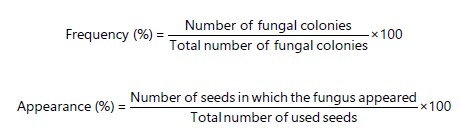
Isolation, identification and purification of fungi associated with cowpea seeds: Random samples were brought from the seeds of two types of cowpea, which were obtained from local markets and the State Company for Seed Production and Certification in Wasit Governorate. The water agar method was used for this, where 50 seeds of each type were superficially sterilized by commercial Sodium Hypochlorite (NaOCl) with a concentration of 2% for 2 min, after which they were washed with sterilized distilled water three times to remove the remnants of the sterilized solution, then the excess water was removed using filter paper. Then, the seeds were transferred by sterilized forceps to Petri dishes containing the nutrient medium PDA, with ten seeds per plate and three replicates for each group, approximately equal distances from each other within one dish. Then the dishes were incubated at a temperature of 25±3°C and after four days, the developing fungal colonies were examined and purified in fresh cultivation medium and the developing fungi were diagnosed to the species level depending on the approved taxonomic characteristics9. The frequency and appearance of the diagnosed fungi were calculated according to the Eq.:
 |
After purification and identification of the isolated fungi from the seeds, they were cultivated on the PDA nutrient medium and placed in 20 mL test tubes in a slanted shape and those tubes were incubated in the incubator at a temperature of 25±3°C for 4 days, after that the tubes were kept in the fridge until use. Considering its renewal in the same manner which is described, whenever it needs.
Pathogenicity of isolated fungi of cowpea seeds on petri dishes: The pathogenicity of the isolated fungi from the seeds of two types of cowpea was tested in petri dishes (9 mm) containing the nutrient medium (PDA) which was prepared as mentioned previously. After hardening, a disc of 1 cm diameter was planted in the center of the dish from each fungus separately and that was from the edge of pure farms aged 5 days and with three replications for each, then the dishes were incubated. The seeds were sterilized with sodium hypochlorite 2% concentration for 2 min, then they were washed with sterilized distilled water several times to remove traces of sterilization, then they were placed on filter paper to remove excess water and then 10 seeds were cultivated inside each dish in a circular motion around the developing colonies for 48 hrs for both fungi and at a distance of 1 cm from the edge of the diametrical growth in a circular motion, then the dishes were incubated again in the incubator and after seven days the percentage of seed germination was calculated10 and according to the following equation:
Use of some biological techniques to resist isolated fungi from the seeds of two types of cowpea biofactor fungus: In this study, an isolate of the biological fungus (T. harzianum) was used and obtained from the biological resistance laboratory, College of Agriculture, University of Kufa by Dr. Azhar Hameed, the teacher at Wasit University, College of Agriculture, which was brought from the Arab Republic of Algeria, where the fungus was grown and propagated on the nutrient medium (PDA) and kept in the fridge at a temperature of 4°C for subsequent laboratory and field studies.
Efficiency test of the biological fungus (T. harzianum) in inhibiting isolated fungi from cowpea: The experiment was made to study the antagonistic ability of (T.harzianum) fungi against pathogenic fungi by using (double culture technique) in petri dishes containing the nutrient medium (PDA) that is sterilized and to test the antagonistic ability of (T. harzianum) fungi against the pathogenic fungi (R. solani), (Alternaria alternate), (A. niger), (F. oxysporum), (Penicillium sp.), the plate was divided into two equal parts and the center of the first section was inoculated with a 0.5 cm diameter disc of pathogenic fungi using a sterilized cork piercing near the edges of the grown colony on the nutrient medium (PDA) at 7 days old as well. As for the center of the second section of the petri dish, it was inoculated with a 0.5 cm disc from the edges of the colony of the biological resistance fungus (T. harzianum) at the age of 7 days. Each treatment was repeated three times. A comparison treatment was also carried out by inoculating the centre of the first section of the dish with pathogenic fungi or the biological resistance fungus only11.
The dishes were placed in an incubator at a temperature of 25±1°C for one week. The antagonistic potential was estimated according to a scale (10) consisting of 5 degrees:
| • | Antagonistic fungus covers the entire dish, including the pathogen | |
| • | Antagonistic fungi cover two-thirds of the dish | |
| • | Antagonistic fungi cover half of the dish | |
| • | Pathogenic fungi cover two thirds of the dish | |
| • | Pathogenic fungi cover the entire dish |
The fungi is considered effective if the degree of antagonism is between (1 and 2). The biological factor is antagonistically effective when it shows a degree of antagonism equal to (2) or less with isolates of pathogenic fungi.
RESULTS AND DISCUSSION
Isolation, identification and purification of fungi associated with cowpea seeds: Table 1 shows the genders and types of fungi that were isolated from the seeds of two cultivars of cowpea plants, as five genders were isolated and identified, most of them were imperfect fungi. The results of the study showed that there was a fluctuation in the total isolates of isolated fungi from seeds, where the highest frequency of the fungus (F. oxysporum) was recorded, reaching 67.9% and a lower frequency value for (Alternaria alternate), which was 50%.
Pathogenicity of isolated fungi from cowpea seeds on petri dishes: All tested fungi made a significant reduction in the percentage of germination of Calendula seeds significantly. Table 2, the percentage of germination reached 10% in the treatment of the fungus (R. solani) and it took first place in the effect on seed germination, followed by the treatment of the fungus (F. oxysporum) at 20%, then the treatment of fungus (A. niger) 33.3% followed by treatment with Penicillium sp., by 53.3%.
The fungus (A. alternate) had the least effect on the germination of Calendula seeds, as the percentage of germination was 60% compared to the control treatment, in which the percentage of germination of Calendula seeds was 90%. The germination of Calendula seeds is affected by the variance between fungi. They may be due to the difference in the pathogenicity between fungi. This means the fungi vary in the severity of their effect on the plants. The genetic structures played important roles in them. The three metabolic activities of microorganisms are important in causing pathogenicity. The pathogenicity is able to make pathogens according to the mechanism of action of these metabolic products9.
The pathogenicity ability of (R. solani) is due to its secretion of many lytic enzymes such as cellulase, pectinase and calctodonase, which work on the breakdown of the cell wall, specifically the middle lamina due to its pectin and cellulose content12.
| Table 1: | Isolated fungi from the seeds of two cultivars of cowpea | |||
| Isolated fungi | First cultivar (white cowpea) |
Second cultivar (red cowpea) |
Total |
| Alternaria alternata | 50 |
0 |
50 |
| Aspergillus niger | 42.8 |
44.4 |
43.6 |
| Fusarium oxysporum | 69.2 |
66.6 |
67.9 |
| Penicillium sp. | 37.5 |
14 |
25.7 |
| Rhizoctonia solani | 15 |
7 |
22 |
| Table 2: | Pathogenicity of some fungi isolated from the seeds of two cultivars of cowpea plant using the seeds of calendula | |||
| Fungi name | Germination percentage of Calendula seeds |
| Alternaria alternata | 60 |
| Aspergillus niger | 33.3 |
| Fusarium oxysporum | 20 |
| Penicillium sp. | 53.3 |
| Rhizoctonia solani | 10 |
| Control | 90 |
| LSD 0.05 | 6.5 |
| Table 3: | Antagonistic ability test of biological resistance fungi (T. harzianum) according to the bell scale with some fungi | |||
| Isolated fungi | Degree of antagonism of the fungus (T.h.) with fungi |
| Alternaria alternata | 3 |
| Aspergillus niger | 4 |
| Fusarium oxysporum | 2 |
| Penicillium sp. | 4 |
| Rhizoctonia solani | 1 |
The fungus (F. oxysporum) is characterized by the fact that it secretes many decomposing enzymes outside and towards the plant host and these enzymes are (Lipase), which increases the pathogenicity ability of the fungus, as well as the mycotoxins that the fungus is characterized by its production13.
Effect of (T. harzianum) in inhibiting the growth of fungi in nutrient medium PDA: The results indicate that the fungus (T. harzianum) had a significant effect in limiting the growth of the pathogenic fungus (R. solani) as it gave an antagonism degree1 based on the scale of Bell 1982. The inhibitory activity of (T. harzianum) against the pathogen was due to its various and well-known mechanisms in controlling pathogens, including competition for food, space and its ability to produce antibiotics and some enzymes that decompose pathogen cell walls such as (protease enzyme), β-1,3 (glucanase) and (chitinase)14. These results were in agreement with the studies of many researchers on the ability of the biological resistance fungus (T. harzianum) to reduce the effect of (R. solani)1, The results of Table 3 indicate a high antagonistic feature of T.h.1, fungi against other fungi. The degree of antagonism was 4 against both (Penicillium sp.) and (A. niger). As for the fungus (oxysporum Fusarium), the degree of antagonism for the bio resistance fungus T.h.1 was 2, while the degree of antagonism with the fungus (A. alternate) was 3 and this may be due to the secretion of antibiotics by the fungus A.a., to defend itself. The results of the experiment showed that the mycelium of the biological resistance fungus T.h.1 grows in all cases above the other mycelium and this agrees with what was found15. From the results of this experiment, it was found that the biological resistance fungus T.h., when antagonized with the fungus R.s., a yellow halo formed in the area where the two fungi meet and it is believed that they are toxic secretions released by the resistance fungus to kill the other fungus and feed on it by a (Necrotrophic) method and this is consistent with what was reached16.
It was mentioned by Neergaard11 that the mechanism of the biological resistance fungus T.h. Through microscopic observations in double cultures, it is found that the mycelium of the biological resistance fungus T.h., is coiling in a spiral shape around the mycelium of the pathogen R.s. The fungus pathogen R.s. secretes materials that are rich in (galactose sugar), which the fungus feeds on, in addition to the fact that the walls of the fungus contain (chitin) in the form of (N-Acetylglucosamine), which is a catalyst for the biological resistance fungus to secrete the enzyme (Chitinase), which leads to the decomposition of the walls of the fungal hyphae of the pathogenic fungus to break down this complex sugar17,18.
CONCLUSION
The study concludes that the fungus (T. harzianum) had a significant effect in limiting the growth of the pathogenic fungus (R. solani). The inhibitory activity of (T. harzianum) against the pathogen was due to its various and well-known mechanisms in controlling pathogens, including competition for food, space and its ability to produce antibiotics and some enzymes that decompose pathogen cell walls such as (Protease).
SIGNIFICANCE STATEMENT
The pathogenicity of fungi isolated from the seeds of two cultivars of cowpea showed that the percentage of germination of Calendula seeds was significant. The germination of Calendula seeds is affected by the variance between fungi. They may be due to the difference in the pathogenicity between fungi. This means the fungi vary in the severity of their effect on the plants. The genetic structures played important roles in that three metabolic activities of microorganisms are important in causing pathogenicity.
REFERENCES
- Thompson, H.C. and W.C. Kelly, 1957. Vegetable Crops. 5th Edn., McGraw Hill, New York, ISBN: 9780070644182, Pages: 611.
- Agrios, G.N., 1997. Plant Pathology. 4th Edn., Academic Press, Cambridge, Massachusetts, ISBN-13: 9780120115648, Pages: 635.
- Domsch, K.H., W. Gams and T.H. Anderson, 1980. Compendium of Soil Fungi. Academic Press, London, United Kingdom, ISBN: 9780122204012, Pages: 1264.
- Bell, D.K., H.D. Wells and C.R. Markham, 1982. In vitro antagonism of Trichoderma species against six fungal plant pathogens. Phytopathology, 72: 379-382.
- Benhamou, N., 1993. Hyphal interactions between Trichoderma harzianum and Rhizoctonia solani: Ultrastructure and gold cytochemistry of the mycoparasitic process. Phytopathology, 83: 1062-1071.
- Bolkan, H.A. and D.F. Butler, 1974. Studies on heterokaryosis and virulence of Rhizoctonia solani. Phytopathology, 64: 513-522.
- Voigt, C.A., W. Schäfer and S. Salomon, 2005. A secreted lipase of Fusarium graminearum is a virulence factor required for infection of cereals. Plant J., 42: 364-375.
- Taylor, R.J., B. Salas, G.A. Secor, V. Rivera and N.C. Gudmestad, 2002. Sensitivity of North American isolates of Phytophthora erythroseptica and Pythium ultimum to mefenoxam (Metalaxyl). Plant Dis., 86: 797-802.
- Khangura, R.K., M.J. Barbetti and M.W. Sweetingham, 1999. Characterization and pathogenicity of Rhizoctonia species on canola. Plant Dis., 83: 699-783.
- Elad, Y., I. Chet, P. Boyle and Y. Henis, 1983. Parasitism of Trichoderma spp. on Rhizoctonia solani and Sclerotium rolfsii-Scanning electron microscopy and fluorescence microscopy. Phytopathology, 73: 85-88.
- Neergaard, P., 1977. Seed Pathology. 2nd Edn., MacMillan, New York, ISBN: 9780333192733, Pages: 1191.
- Nazeer, W.W., 2022. A novel isolation and purification of antifungal chitinase from cowpea (Vigna unguiculata) and its possible biotechnological applications. Afr. J. Biol. Sci., 18: 89-96.
- de Beeck, M.O., P. Persson and A. Tunlid, 2021. Fungal extracellular polymeric substance matrices-Highly specialized microenvironments that allow fungi to control soil organic matter decomposition reactions. Soil Biol. Biochem., 159.
- Haleem, R.A., K.A. Saedo and S.K. Abdullah, 2016. Antagonism of Trichoderma harzianum and Clonostachys rosea against fungi associated with grapevine decline in Kurdistan Region-Iraq. Sci. J. Univ. Zakho, 4: 166-172.
- Ouili, A.S., Y. Maiga, M. Nikiema, S. Bissiri and Y. Dabiré et al., 2023. Post-harvest fungi associated with cowpea (Vigna unguiculata L. Walp.) seeds produced in Burkina Faso. Adv. Microbiol., 13: 148-163.
- Siddiquee, S., F. Abdullah, T.S. Guan and L.M. See, 2007. Allozyme variations of Trichoderma harzianum and its taxonomic implications. Aust. J. Basic Appl. Sci., 1: 30-37.
- Gowda, B.S., J.L. Miller, S.S. Rubin, D.R. Sharma and M.P. Timko, 2002. Isolation, sequence analysis, and linkage mapping of resistance-gene analogs in cowpea (Vigna unguiculata L. Walp.). Euphytica, 126: 365-377.
- Whipps, J.M., 1997. Developments in the biological control of soil-borne plant pathogens. Adv. Botanical Res., 26: 1-134.
How to Cite this paper?
APA-7 Style
Tofan,
A.J., Khiry,
S.B. (2024). Isolation, Purification and Identification of Fungi Associated with Cowpea Seeds, Vigna unguiculata (L.) and the Possibility of Their Resistance Using Some Biological Techniques. Asian Journal of Biological Sciences, 17(3), 284-290. https://doi.org/10.3923/ajbs.2024.284.290
ACS Style
Tofan,
A.J.; Khiry,
S.B. Isolation, Purification and Identification of Fungi Associated with Cowpea Seeds, Vigna unguiculata (L.) and the Possibility of Their Resistance Using Some Biological Techniques. Asian J. Biol. Sci 2024, 17, 284-290. https://doi.org/10.3923/ajbs.2024.284.290
AMA Style
Tofan
AJ, Khiry
SB. Isolation, Purification and Identification of Fungi Associated with Cowpea Seeds, Vigna unguiculata (L.) and the Possibility of Their Resistance Using Some Biological Techniques. Asian Journal of Biological Sciences. 2024; 17(3): 284-290. https://doi.org/10.3923/ajbs.2024.284.290
Chicago/Turabian Style
Tofan, Amir, Jassim, and Suadad Breesam Khiry.
2024. "Isolation, Purification and Identification of Fungi Associated with Cowpea Seeds, Vigna unguiculata (L.) and the Possibility of Their Resistance Using Some Biological Techniques" Asian Journal of Biological Sciences 17, no. 3: 284-290. https://doi.org/10.3923/ajbs.2024.284.290

This work is licensed under a Creative Commons Attribution 4.0 International License.