Evaluation of Anti-Ulcerogenic Effects of the Crude Extract and Fractions of Dialium guineense Tree Bark in Ethanol-Induced Peptic Ulcer in Albino Rats
-
Obidike Ikechukwu Johnlouis


Department of Biochemistry, College of Natural Sciences, Michael Okpara University of Agriculture, Umudike, Nigeria
Aloh Godwin SundayDepartment of Biochemistry, College of Natural Sciences, Michael Okpara University of Agriculture, Umudike, Nigeria
Obike Chiemeziem AdanmaDepartment of Biochemistry, College of Natural Sciences, Michael Okpara University of Agriculture, Umudike, Nigeria
Okechukwu Jane NgoziDepartment of Biochemistry, College of Natural Sciences, Michael Okpara University of Agriculture, Umudike, Nigeria
| Received 29 Nov, 2023 |
Accepted 15 Feb, 2024 |
Published 30 Sep, 2024 |
Background and Objective: Medicinal plants contain bioactive molecules with pharmacological potential for managing diseases. Peptic ulcer is a ravaging disease that has been attributed to cases of mortality across the globe and Dialium guineense; a medicinal plant could form bases for its amelioration hence, this study sets out to evaluate the anti-ulcerogenic potentials of a crude ethanol extract and fractions of Dialium guineense stem bark in ethanol-induced ulcer in albino rats. Materials and Methods: The biochemical effects of Dialium guineense stem bark in ethanol-induced ulcers were determined using standard protocols. The SPSS version 22 was used for the one-way ANOVA analysis (p<0.05). Results: For the gastro-protective estimation, ulcer index and percentage protection score of the treatment groups were significantly (p<0.05) lower than those of the untreated groups and the standard drug (omeprazole) in a dose-dependent manner. There were significant (p<0.05) elevations in red blood cell and hemoglobin concentrations respectively in the groups co-treated with different fractions of Dialium guineense. The result also indicated a significant (p<0.05) decrease in the tumor necrotic factor-α and the interleukin-1β concentration across the fractions group compared with the normal group. The result of the antioxidant study showed significant (p<0.05) increases in the superoxide dismutase, catalase activities and glutathione concentrations of the co-treated groups compared to the untreated group. The organ histology showed protected stomach architecture in the co-treated groups compared to that of the negative control which showed full laceration of the stomach wall. Conclusion: Hence, it could be concluded that different fractions of Dialium guineense stem bark extract have an organo-protective activity which could be attributed to its high antioxidant activity as antioxidants have been found to reverse the toxicity caused by pro-oxidants.
| Copyright © 2024 Johnlouis et al. This is an open-access article distributed under the Creative Commons Attribution License, which permits unrestricted use, distribution, and reproduction in any medium, provided the original work is properly cited. |
INTRODUCTION
From time immemorial, parts of medicinal plants and their ingredients have been used as primary therapeutic agents in the management of disease conditions1-3. For centuries, medicinal herbaceous plants including Polygonum chinense L.4, Euphorbia hirta L.5 and Ilex rotunda Thunb6 have been and are being processed into herbal tea for the management of such disease conditions as dyspepsia, enteritis, gastric ulcer and other digestive system diseases.
Dialium guineense (DAG), an indigenous African plant, has been demonstrated to be useful in the treatment of peptic ulcers by local users, particularly when taken as a tea7. The leaves especially have been shown to possess this activity8. Dialium guineense (Wild) belongs to the family of Fabaceae, commonly referred to as “black velvet” or “velvet tamarind”9. It is a tree plant with an average height of 30 m densely hairy leaves and smooth greyish bark. Flowers are often pale and fruits are red usually flattened and circular10.
Peptic ulcer is a disease usually referred to as Peptic Ulcer Disease (PUD). It is a defect in the gastrointestinal mucosa that extends across the mucosae muscularis and occurs as a result of the acidic or peptic activity in the gastric juice11. In Nigeria, the prevalence of PUD is not fully known possibly due to people resorting to self-medication using local means12. However, the complications attributed to PUD have led to high mortality rates with perforations contributing about 30% hemorrhage (about 10%); while obstruction is the commonest complication (56%)13.
Two aetiological factors: The use of Nonsteroidal Anti-Inflammatory Drugs (NSAIDs) and Helicobacter pylori infection have been largely implicated in the pathogenesis of peptic ulcer disease14. Over time, the incidence of H. pylori-mediated duodenal ulcers in the developed world has become much less than that of the developing countries because of decrease in the intake of NSAIDs in the former14.
Excessive intake of alcohol has been shown to be one of the main causes of acute gastric mucosal damage in humans15; for this reason absolute ethanol administration is usually used to induce gastric injury in animal models, including rats16. Administration of highly concentrated ethanol provokes oxidative stress and subsequently induces inflammation as a mechanism for the pathogenesis of gastric injury17. This is because ethanol causes the production of reactive oxygen species that cause depletion of cellular antioxidant enzymes which, in turn, leads to the induction of oxidative stress in many ways18. Oxidative stress then leads to the production of numerous pro-inflammatory mediators19 with subsequent inflammatory response20. Consequently, inflammatory reactions produce reactive oxygen species which cause oxidative stress that ultimately leads to ulcerative damage21. Consequent to frequent consumption of alcohol by people of all ages for various reasons, including socialisation and reports of Dialium guineense being useful in the traditional management of ulcers, this study is aimed at establishing a scientific basis for the folkloric use of the plant in the management of alcohol-induced ulcer in albino rats.
MATERIALS AND METHODS
Collection and identification of sample: The study took place in the laboratory unit of Michael Okpara University of Agriculture, Umudike Abia State, Nigeria between April, 2023 to August, 2023. Dialium guineense stem bark was obtained from Ozubulu in Ekwusigo Local Government Area of Anambra State. The plant was identified and authenticated by Prof. G.G.E. Osuagwu of the Department of plant Science and Biotechnology, Michael Okpara University of Agriculture Umudike, Abia State, Nigeria. A voucher specimen was kept at the herbarium in the Department of Physiology and Pharmacology, College of Veterinary Medicine.
Preparation of extract: The Dialium guineense bark was thoroughly washed with tap water to avoid contamination of samples by dirt. The washed sample was then air-dried at room temperature for eight weeks. The dried plant sample was then milled using a locally made hard metal grinder. Fifty grams of the powdered sample was extracted using 96% ethanol in a Soxhlet extractor (model SE-6P Infitek, China). Temperature for extraction was maintained at 60°C for 72 hrs. The dried extract was then oven-dried (IEC60068-2-2 Dongguan China) at 40°C to obtain a dark yellow semi-solid sample that weighed 2.00 g. The crude ethanol extract was stored in a refrigerator (Thermocool, Nigeria) at 10°C until the time of use. Percentage yield of the extract was calculated using the formula22:
Where:
X = Weight of dried Dialium guineense bark extract after extraction (2.00 g)
Q = Weight of Dialium guineense bark extract powdered plant material before extraction
Study design study design for the biochemical estimation of Dialium guineense bark extract: Thirty male albino rats were obtained from the animal house of Michael Okpara University, Umudike. The animals were randomly grouped into 6 groups, placed in an aluminum cage containing 5 animals each and acclimatized for 12 days. All of the animals were kept in standard laboratory settings and fed ad libitum with 12 hrs light and dark cycles at room temperature. Every rule pertaining to the use and maintenance of lab animals was strictly followed.
The animals were grouped as such:
• |
Group 1: Normal control (received food and water only) ad libitum |
|
• |
Group 2: Negative control (received 1 mL of ethanol only) |
|
• |
Group 3: Drug control (received 1 mL of ethanol+omeprazole 20 mg) |
|
• |
Group 4: Ethanol group (received 250 mg/bw of ethanol fraction extract+1 mL of ethanol) |
|
• |
Group 5: Methanol group (received 250 mg/bw of methanol fraction extract+1 mL of ethanol) |
|
• |
Group 6: N-hexane group (received 250 mg/bw of N-hexane fraction extract+1 mL of ethanol) |
The plant extract was administered orally to the animals based on their body weights with the aid of an oral gavage and the ethanol was administered 2 hrs later for 21 days. At the end of the administration, the animals were sacrificed through cervical dislocation and blood samples were collected via cardiac puncture while organs of the rats were collected for organ histology.
Determination of gastric ulcer index (UI) and dipercentage protection: The method described by Nwafor et al.23 was used. The extent of damage to gastric mucosa was estimated to determine gross pathology and graded for severity of gastric lesions in a scoring system of 0-5 as proposed by Nwafor et al.23: No lesions (0); small round hemorrhagic lesions (1); lesions less than 2 mm (2); lesions of 2-3 mm (3); lesions 3-4 mm (4) and lesion greater than 4 mm (5). For lesions with a width above 1 mm, the score was multiplied by 2. The percentage of inhibition was calculated by the following formula:
Area of gastric hemorrhagic ulcerative lesions: The area was estimated using the method described by Nwafor et al.23 was used. The dissected rat’s stomach was thoroughly rinsed with cold saline to remove any contaminants and then photographed. The digital photos were used for the estimation degree of gastric lesion (mm2) using ImageJ 1.48d software. The gastric lesions seen were recorded including the multifocal erosions and the linear hemorrhagic lesions.
Examination and microscopic scoring of gastric damage: The estimation of gastric damage was done by the method described by Budimi et al.24. Gastric layers were fixed in 10% buffered formol saline for 24 hrs. The tissue sample was then washed, dehydrated by alcohol and embedded in paraffin in a hot air oven (56°C) for another 24 hrs. The layer was sliced to 5 μm thickness, stained with H&E and examined under the light microscope (Leica Microsystems, Germany).
Evaluation of antioxidant enzymes activities
Catalase: The catalase activity was evaluated by the method described by Obidike and Ngozi25, based on the principle that catalase splits hydrogen peroxide. At specific time intervals, the reaction was stopped by the addition of a dichromate/acetic acid reagent. When heated, hydrogen peroxide reacts with dichromate in acetic acid to form reduced chromic acetate and an unstable perchromic intermediate. The excess hydrogen peroxide is then determined by measuring the optical density of chromic acetate formed at a wavelength of 610 nm.
Reagent composition: The chemical composition of the reagent includes phosphate buffer (0.05M pH 7.4).
Standard hydrogen peroxide (0.2M), dichromate/acetic acid reagent, stock (5% potassium dichromate in glacial acetic acid 1:3) and working dichromate reagent (1 mL stock diluted with 4 mL water).
Assay mixture was composed of hydrogen peroxide, 1.0 mL of buffer, 0.4 mL of distilled water and 0.1 mL of dilute organ homogenate (1:10). The reaction stopped after a period of 60 seconds of incubation. As 20 mL of dichromate/acetic acid reagent was added to stop the reaction. The serum was then introduced into the control tube after the addition of a reagent. The tubes were heated in a water bath for 10 min and the colour developed was read at 610 nm. Various concentrations of the standard within the range of 20-120 moles were used to obtain the calibration curve. The activity of the catalase was determined by the amount of hydrogen peroxide consumed expressed in moles of H2O2 consumed/minute/mL.
Evaluation of superoxide dismutase (SOD) activity: The method of auto-oxidation by pyrogallol as described by Obidike and Ngozi25 was used.
Principle: Pyrogallol auto oxidizes rapidly in an alkaline solution to generate superoxide ions. The SOD inhibits its auto oxidation dismutating the superoxide ions to hydrogen peroxide and molecular oxygen. The activity of 50% inhibition by SOD was measured at 450 nm.
Reagent composition: The chemical composition of the reagent includes Tris-HCL (0.55M, pH 7.4), buffer (Tris-HCL 0.05 M, 1 mM EDTA, pH 8.5), pyrogallol stock (24.2 mg of pyrogallol in 1 mL of Tris-HCL dilution ratio 1:1000), absolute ethanol and chloroform.
Procedure: Absolute ethanol (0.25 mL) and 0.1 mL of chloroform were added to cuvette and vigorously shaken and centrifuged for 30 min at 1000 rpm. The supernatant was used for the assay. About 0.5 mL of the stock solution of pyrogallol was diluted to 50 mL with Tris-HCl with a final concentration of 0.2 mM. The assay mixture for autooxidation without the enzyme source was composed of 2.0 mL buffer containing EDTA and 0.5 mL of diluted pyrogallol and water to bring the final volume to 3.0 mL and the OD read initially after 3 min at 420 nm. The assay mixture for oxidation with enzymes was composed of 2.0 mL buffer solution containing EDTA 0.5 mL pyrogallol and 0.1 mL of liver supernatant bringing the total volume to 3.0 mL with distilled water. The absorbance was also read at the same wavelength (420 nm).
Evaluation of glutathione reductase activity: The method described by Obidike and Ngozi25 was used for the determination of glutathione.
Principle: It involves the development of a yellow colour using Ellman’s reagent. The 5,5, Dithio-bis-2-Nitrobenzoic Acid (DTNB) to compounds containing sulphydryl groups. The absorbance was read at 412 nm.
Reagent composition: The reagent consisted of chemical composition of the reagent including DTNB (0.1 M methanol), TCA (trichloro acetate) (5%), phosphate buffer (0.05 M, pH 7.4), EDTA (1.0 mM) and standard (reduced glutathione 10 mg in 100 mL of distilled water).
Procedure: To 0.1 mL of the homogenate, 2.4 mL of EDTA, 0.1 mL distilled water and 1.5 mL TCA were added and mixed thoroughly. The mixture was centrifuged after 5 minutes to obtain the supernatant. To 0.1 mL of the supernatant, 1.0 mL of DTNB and 1.0 mL of buffer were added. The absorbance was read at 412 nm against reagent blank. The result was expressed in μmol/mg protein.
Evaluation of haematological indices: The selected haematological indices were evaluated by the standard method described by Johnlouis et al.26. The indices examined were RBC, WBC, PCV and Hb. The standard procedures used were as follows.
Determination of blood cell counts (WBC and RBC)
Principle: This was obtained using an improved Neubauer chamber microscopic grid scoring platform in which the specific type and quantity of blood fluid were retained. By properly diluting the blood, counting the squares and multiplying by the right conversion factor (square indicated under magnification objective lens, the number of each cell per cubic millimetre of blood can be estimated). The number of blood cells in a cubic millimetre of blood can be estimated by diluting a sample of blood using appropriate fluid into a glass containing the cells in a known volume. A sample calculation provides the number of cells in 1 mm of blood using the improved Neubauer chamber. With the aid of appropriate diluents and dilutions, the number of red cells, white cells and platelets can be determined.
Diluents include; Hayem’s solution for RBC and 1% HCl for WBC.
Procedure: As 0.02 mL of blood was added to 4 mL of diluents in a test tube (1:200). The test tubes were sealed and the content was allowed to mix for 1 min. The counting chambers were filled by means of the capillary tube and counted after 2-5 min in 1/5 square indicated under ×10 magnification objective lens.
Calculation: Johnlouis et al.26:
 |
Measurement of Packed Cell Volume (PCV)
Principle: The PCV was determined by microhematocrit method as described by Johnlouis et al.26. The volume occupied by the red blood cell in a given volume of whole blood expressed as a percentage is PCV.
Procedure: The capillary tubes containing the blood samples to which EDTA anticoagulant was added were sealed with plasticine and centrifuged for 5 min at 10,000 rpm to give three suspensions: Grey layer (mass of erythrocytes), thrombocytes (buffy coat) and plasma from bottom to top in a hematocrit reader on which the percentage of PCV was read off.
Calculation: Johnlouis et al.26:
 |
Evaluation of Tumor Necrosis Factor (TNF-α)
Pro-inflammatory cytokine assay
Tumor Necrosis Factor-Alpha (TNF-α): An ELISA-based method was used for the assay.
Principle: The ELISA kit uses sandwich-ELISA as the method. The micro-ELISA plate provided in the kit was precoated with an antibody-specific rat TNF-α. Standards or samples were added to appropriate micro-ELISA plate wells and combined with the specific antibodies. Then biotinylated detection antibodies specific for rat TNF-α and Avidin horseradish peroxidase (HPR) conjugate were added to each microplate well and incubated. Free components in the wells were washed using diluted wash reagent. Then the substrate reagent was added to each well. Only wells containing rat TNF-α, biotinylated detection antibody and Avidin HRP conjugate developed a blue colour. The enzyme-substrate reaction was terminated by adding the stop solution and the reaction stopped when a yellow colour developed. The optical density (OD) was then measured using a spectrophotometer at a wavelength of 450 nm. The OD value is proportional to rat TNF-α concentration.
Test preparations: Tissue homogenate of rat hearts was used. The heart tissues were washed with 0.01 M phosphate buffer solution before homogenization. The tissue homogenate was then mixed with the same concentration of buffer in the ratio of 1:9 (tissue weight: volume of buffer). Homogenate was then centrifuged for 5 min at 500 nm to obtain the supernatant. Samples and reagents were all brought to room temperature before the analysis. A volume (1 mL) of stock wash buffer solution was measured out and made up to 750 mL of a working buffer solution using deionised water.
Standard working solution: Stock standard solution provided was first centrifuged at 10,000 rpm for 1 min. One milliliter of stock and 1 mL of sample diluents were measured into test tubes and mixed by inverting the tubes and later incubating for 10 min. This gave a standard solution of 500 mg/mL. Serial dilutions were then made as follows: 5,000, 2,500, 1,250, 625, 312.5, 156.25, 78.13 and 0 pg/mL.
Biotinylated detection antibody working solution: The 100X concentrated biotinylated detection antibody was diluted to 1X working solution with biotinylated detection antibody Ab diluents.
Concentrated HRP conjugate working solution: The 100X concentrated HRP conjugate was diluted to 1x working solution with HRP conjugate diluents.
Procedure:
• |
The 100 μL of standard or sample was added to each of either standard or sample and incubated for 90 min at 37°C |
|
• |
Solution was removed and 100 μL biotinylated detection Ab was added and incubated for 1 hr at 37°C |
|
• |
Resulting solution was then aspirated and washed 3 times using the wash solution |
|
• |
The 100 μL conjugate was added and incubated for 30 min at 37°C |
|
• |
Resulting solution was aspirated and washed 5 times |
|
• |
90 μL stop solution was added and OD was read immediately using a microplate reader at 450 nm |
|
• |
A graph of OD was plotted against concentration for known standard concentration and concentration for sample extrapolated from a graph |
Evaluation of interleukin 1 beta (IL-1β) concentration: An ELISA-based method was employed for the assay. The kit was manufactured by Elabscience and the instructions stated on it were adhered to strictly.
Principle: The micro-ELISA plate provided was pre-coated with an antibody specific to IL-1β. Standards or samples were added to appropriate micro ELISA plate walls and combined with specific antibodies. Then the biotinylated detection antibody specific for rat IL-1β and Avidin horseradish peroxidase conjugate were added to each microplate and incubated. After incubation, the resulting complex was washed using the wash reagent. Then the substrate reagents were added to each well. Only the wells containing rat IL-1β, biotinylated antibody and Avidin horseradish peroxidase will develop blue colour. The enzyme-substrate reaction is terminated by adding a stop solution to develop a yellow colour. The optical density (OD) is immediately measured using a spectrophotometer (Labtech, America Model Spectronic-20) at 450 nm. The OD value is proportional to the concentration of rat IL-1β.
Reagent preparation: Tissue homogenates of rats’ hearts were used. Heart tissues were washed with 0.01M phosphate buffer solution before homogenization. The tissue homogenate was then mixed with the same concentration of buffer in the ratio 1:9 (tissue weight:volume of buffer). Homogenate was then centrifuged for 5 min at 500 rpm to obtain the supernatant. Samples and reagents were all brought to room temperature before analysis.
Wash buffer: As 300 mL of stock wash buffer solution was measured out and made up of 750 mL of working buffer solution with deionized water.
Standard working solution: Stock standard solution was first centrifuged at 10,000 rpm for 1 min. As 1 mL of stock solution and 1 mL of sample diluents were measured into the test tube and mixed by inverting the test tubes and later incubating for 10 min. This gave a standard working solution of 500 pg/mL. Serial solutions were then made in the gradient as follows 500, 250, 125, 62.5, 31.5, 15.63, 7.81 and 0 Pg/mL.
Biotinylated detection antibody working solution: The 100X concentrated biotinylated detection Ab was diluted to 1X working solution using biotinylated detection Ab diluents.
Concentrated horseradish peroxidase (HRP) conjugate working solution: The 100X concentrated HRP conjugate was diluted to 1X working solution using HRP conjugate diluents.
Procedure: As 100 μL standard of sample was added to each well and incubated for 90 min at 37°C. Liquid was removed and 100 μL biotinylated detection Ab was added and incubated for 1 hr at 37°C. The solution was inspired and well-washed 3 times. As 100 μL HRP conjugate was added and incubated for 30 min at 37°C. The solution was aspirated and washed 5 times. As 90 μL of substrate reagent was added and incubated for 15 min at 37°C.
As 50 μL of stop solution was then added and optical density was immediately taken using a microplate reader at 450 nm. Graph of OD plotted against concentration for known standard and concentration for sample for known standard and concentration for sample extrapolated in the graph. Duplicate readings were encouraged.
RESULTS
Table 1 shows gastroprotective effects of different fractions of Dialium guineense stem bark in ethanol-induced ulcers in albino rats.
| Table 1: | Gastroprotective effects of different fractions of Dialium guineense stem bark in ethanol-induced ulcer in albino rats | |||
| Groups | Ulcer score |
Average number of ulcer per animal |
Animal with ulcer (%) |
Ulcer index |
Protection (%) |
Gastric pH |
| Group 1 (normal group) | 0* |
0 |
0 |
0* |
100* |
3.04* |
| Group 2 (negative control) | 24 |
11 |
100 |
13 |
0 |
2.6 |
| Group 3 (drug control) | 16* |
7 |
80 |
10* |
25.25* |
3.31* |
| Group 4 (ethanol 250 mg/kg extract+1 mL ethanol) | 7* |
4 |
80 |
9* |
33.54* |
3.63* |
| Group 5 (methanol 250 mg/kg extract+1 mL methanol) | 4* |
2 |
82 |
8* |
38.39* |
3.71* |
| Group 6 (N-hexane 250 mg/kg extract+1 mL ethanol) | 2* |
2 |
70 |
7* |
48.83* |
3.85 |
| Values are mean and *Statistically significant (p<0.05) compared to negative control (group 2) | ||||||
| Table 2: | Effects of fractions of Dialium guineense bark extract on serum SOD and catalase activity of ethanol-induced ulcer in Wistar albino rats | |||
| Groups | SOD (μmol/L) |
CAT (μmol/L) |
| Group 1 (normal group) | 39.36±1.30 |
21.42±2.50 |
| Group 2 (negative control) | 34.58±2.23 |
18.40±1.35 |
| Group 3 (drug control) | 34.56±1.19 |
19.46±0.87* |
| Group 4 (ethanol 250 mg/kg extract+1 mL ethanol) | 35.56±0.56 |
18.96±0.87 |
| Group 5 (methanol 250 mg/kg extract+1 mL ethanol) | 36.96±0.08* |
19.60±1.40* |
| Group 6 (N-hexane 250 mg/kg extract+1 mL ethanol) | 37.62±0.51* |
19.84±2.15* |
| Values are Mean±Standard Deviations and *Statistically significant (p<0.05) compared to negative control (group 2) | ||
The ulcer score count suggested that the counts of the negative control were significantly (p<0.05) higher than that of the normal group while the count of the co-treated groups was significantly (p<0.05) lower when compared with that of the negative control.
The average number of ulcer counts per animal was highest in the negative control group while those of the drug and the co-treated groups were lower compared when to those of the negative control.
For the ulcer index estimation, the mean index of the negative control group was significantly (p<0.05) higher than that of the normal control. However, the index of the drug control and those of the co-treated groups were significantly (p<0.05) lower than that of the negative control.
The percentage protection of the negative control was zero, thus significantly (p<0.05) lower than that of the normal control which had 100% protection. However, the percentage protection of the drug control group (25.25%) and those of the co-treated groups were significantly (p<0.05) lower than that of the negative control.
The pH of the negative control was also significantly (p<0.05) lower than that of the normal group while the pH of the drug control group and those of the groups that received different concentrations of Dialium guineense bark extract were all significantly (p<0.05) lower than that of the negative control.
Table 2 shows the effects of fractions of Dialium guineense bark extract on SOD and catalase activity of ethanol-induced ulcer in Wistar albino rats.
The result of the SOD activity showed that the activity of group 3 (drug group) was the lowest followed closely by that of group 2 (negative control). The SOD activity of the co-treated groups was higher than those of both groups 2 and 3.
The catalase activity result showed that group 2 (negative control) activity was the lowest, followed by group 3 (drug control). The activity of the co-treated groups was higher than that of the negative and the drug control group.
| Table 3: | Effects of Dialium guineense stem bark methanol extract on serum GSH and MDA concentration of ethanol induced ulcer in Wistar albino rats | |||
| Groups | GSH (μmol/L) |
MDA (μmol/L) |
| Group 1 (normal group) | 55.44±2.43 |
0.30±0.05 |
| Group 2 (negative control) | 41.98±1.88 |
0.78±0.05 |
| Group 3 (drug control) | 47.14±1.74* |
0.70±0.03 |
| Group 4 (ethanol 250 mg/kg extract+1 mL ethanol) | 48.44±1.47* |
0.66±0.05* |
| Group 5 (methanol 250 mg/kg extract+1 mL ethanol) | 52.42±1.49* |
0.59±0.03* |
| Group 6 (N-hexane 250 mg/kg extract+1 mL ethanol) | 54.04±3.07* |
0.52±0.04* |
| Values are Mean±Standard Deviations and *Statistically significant (p<0.05) compared to negative control (group 2) | ||
| Table 4: | Effects of different fractions of Dialium guineense stem bark on RBC, PCV and Hb count in ethanol induced ulcer in albino rats | |||
| Groups | RBC (1012/L) |
PCV (%) |
Hb (g/L) |
| Group 1 (normal group) | 6.80±0.26* |
43.4±2.51* |
16.14±0.52* |
| Group 2 (negative control) | 6.65±0.06 |
41.4±0.89 |
15.92±0.29 |
| Group 3 (drug control) | 6.35±0.26 |
40.2±2.17 |
15.48±0.41 |
| Group 4 (ethanol 250 mg/kg extract+1 mL ethanol) | 6.53±0.43 |
41.4±3.21 |
15.58±0.49 |
| Group 5 (methanol 250 mg/kg extract+1 mL ethanol) | 7.22±0.31* |
46.2±1.30* |
16.72±0.58* |
| Group 6 (N-hexane 250 mg/kg extract+1 mL ethanol) | 7.42±0.05* |
48.8±0.84* |
17.36±0.50* |
| Values are Mean±Standard Deviations and *Statistically significant (p<0.05) compared to negative control (group 2) | |||
Table 3 shows the effects of different fractions of Dialium guineense stem bark on the GSH and MDA concentration in ethanol-induced ulcers in albino rats.
The result of the GSH concentration indicated that the mean concentration of group 2 (negative control) was the lowest followed by group 3 (drug control) while the mean concentrations of the co-treated groups were relatively higher than those of groups 2 and 3.
The results of the MDA assay showed that the mean concentration of group 2 rats (negative control) was the highest followed by group 3 (drug control). However, the mean concentrations of the co-treated groups were lower than those of the groups 2 and 3.
Table 4 shows the effects of different fractions of Dialium guineense stem bark on RBC, PCV and HB count in ethanol-induced ulcers in albino rats.
The result of the RBC count indicated that the count of the negative group was significantly (p<0.05) lower than that of the control group. However, the counts for the groups that received different fractions were significantly (p<0.05) higher than those of the negative control.
The results of the PCV and Hb counts show that those of the negative group (group 2) were significantly (p<0.05) lower than those of the control group, while those of the fractions were significantly (p<0.05) higher than those of the negative control.
Table 5 shows the effects of different fractions of Dialium guineense stem bark on TNF-α and IL-1β in ethanol-induced ulcer in albino rats.
The result of the TNF-α analysis indicated that the mean TNF-α concentration of the group 2 (negative control) rats was significantly (p<0.05) elevated compared with that of the normal control. However, the concentration recorded for different fractions was significantly (p<0.05) reduced compared with those of the untreated groups.
The result of the IL-1β indicated that the mean IL-1β concentration of the group 2 (negative control) rats was significantly (p<0.05) elevated compared with that of the control while those obtained with the different fractions were significantly (p<0.05) reduced in comparison with that of the untreated group.
| Table 5: | Effects of different fractions of Dialium guineense stem bark on TNF-α and IL-1β in ethanol-induced ulcers in albino rats | |||
| Groups | TNF-α (pg/mL) |
IL-1β (pg/mL) |
| Group 1 (normal group) | 59.82±0.84* |
3.60±0.10* |
| Group 2 (negative control) | 70.00±1.20 |
4.10±0.18 |
| Group 3 (drug control) | 67.48±2.29 |
3.75±0.11 |
| Group 4 (ethanol 250 mg/kg extract+1 mL ethanol) | 64.96±3.12* |
3.04±0.09* |
| Group 5 (methanol 250 mg/kg extract+1 mL ethanol) | 66.30±1.86* |
3.63±0.39* |
| Group 6 (N-hexane 250 mg/kg extract+1 mL ethanol) | 63.48±0.88* |
3.22±0.10* |
| Values are Mean±Standard Deviations and *Statistically significant (p<0.05) compared to negative control (group 2) | ||
| Table 6: | Effects of different fractions of Dialium guineense stem bark on PGE2 concentration in ethanol-induced ulcer in albino rats | |||
| Groups | PGE2 (pg/mL) |
| Group 1 (normal group) | 28.87±0.54 |
| Group 2 (negative control) | 52.16±0.69 |
| Group 3 (drug control) | 44.10±0.60* |
| Group 4 (ethanol 250 mg/kg extract+1 mL ethanol) | 43.17±0.39* |
| Group 5 (methanol 250 mg/kg extract+1 mL ethanol) | 40.2±0.82* |
| Group 6 (N-hexane 250 mg/kg extract+1 mL ethanol) | 36.94±0.43* |
| Values are Mean±Standard Deviations and *Statistically significant (p<0.05) compared to negative control (group 2) | |
| Table 7: | Effects of different fractions of Dialium guineense stem bark on LDH concentration in ethanol-induced ulcer in albino rats | |||
| Groups | LDH (U/L) |
| Group 1 (normal group) | 289.23±0.56 |
| Group 2 (negative control) | 423.69±0.69 |
| Group 3 (drug control) | 344.22±0.65* |
| Group 4 (ethanol 250 mg/kg extract+1 mL ethanol) | 333.17±0.90* |
| Group 5 (methanol 250 mg/kg extract+1 mL ethanol) | 340.20±0.24* |
| Group 6 (N-hexane 250 mg/kg extract+1 mL ethanol) | 387.94±0.37* |
| Values are Mean±Standard Deviations and *Statistically significant (p<0.05) compared to negative control (group 2) | |
Table 6 shows the effects of different fractions of Dialium guineense stem bark on PGE2 concentration in ethanol-induced ulcers in albino rats.
The result of the PGE2 estimation showed a significant (p<0.05) increase in serum concentration of the untreated group compared with that of the normal group. However, the PGE2 serum concentration of the co-treated groups all showed a significant (p<0.05) decrease compared with that of the untreated group.
Table 7 shows the effects of different fractions of Dialium guineense stem bark on LDH concentration in ethanol-induced ulcers in albino rats.
The result of the LDH estimation indicated a significantly (p<0.05) increased serum concentration in the untreated group 2 compared with that of the normal group. However, the serum concentrations for the co-treated groups all indicated significantly (p<0.05) decreased concentration compared with that of the untreated group even lower than that of the standard treatment drug.
Table 8 shows the effects of different fractions of Dialium guineense stem bark on CPK concentration in ethanol-induced ulcers in albino rats.
The result of the CPK estimation indicated a significant (p<0.05) increase in serum concentration compared to that of the normal group. However, the CPK serum concentration in the co-treated groups indicated significant (p<0.05) reduced CPK compared with that of the untreated group even lower than that of standard treatment drug.
| Table 8: | Effects of different fractions of Dialium guineense stem bark on CPK concentration in ethanol-induced ulcer in albino rats | |||
| Groups | CPK (U/L) |
| Group 1 (normal group) | 200.70±0.10 |
| Group 2 (negative control) | 240.36±0.23 |
| Group 3 (drug control) | 225.70±0.70* |
| Group 4 (ethanol 250 mg/kg extract+1 mL ethanol) | 217.20±0.10* |
| Group 5 (methanol 250 mg/kg extract+1 mL ethanol) | 215.20±0.24* |
| Group 6 (N-hexane 250 mg/kg extract+1 mL ethanol) | 230.24±0.99* |
| Values are Mean±Standard Deviations and *Statistically significant (p<0.05) compared to negative control (group 2) | |
| Table 9: | Effects of different fractions of Dialium guineense stem bark on troponin concentration in ethanol-induced ulcer in albino rats | |||
| Groups | Troponin |
| Group 1 (normal group) | 60.70±0.33 |
| Group 2 (negative control) | 73.67±0.70 |
| Group 3 (drug control) | 71.70±0.53 |
| Group 4 (ethanol 250 mg/kg extract+1 mL ethanol) | 70.21±0.67* |
| Group 5 (methanol 250 mg/kg extract+1 mL ethanol) | 65.20±0.33* |
| Group 6 (N-hexane 250 mg/kg extract+1 mL ethanol) | 70.33±0.33* |
| Values are Mean±Standard Deviations and *Statistically significant (p<0.05) compared to negative control (group 2) | |
Table 9 shows the effects of different fractions of Dialium guineense stem bark on troponin concentration in ethanol induced ulcer in albino rats.
The result of the cardiac troponin estimation indicated a significant (p<0.05) increase in serum concentration of the untreated group compared with that of the normal group. However, the serum concentration of troponin estimation in the co-treated groups showed a significant (p<0.05) decrease compared with that of the untreated group.
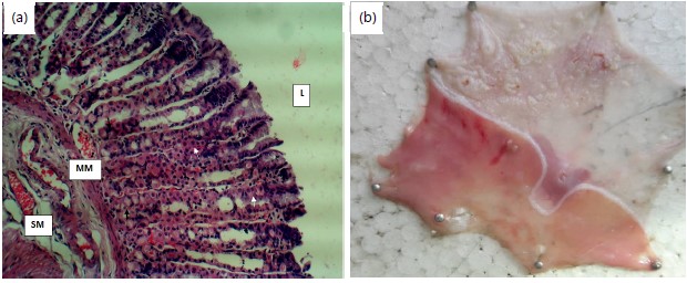
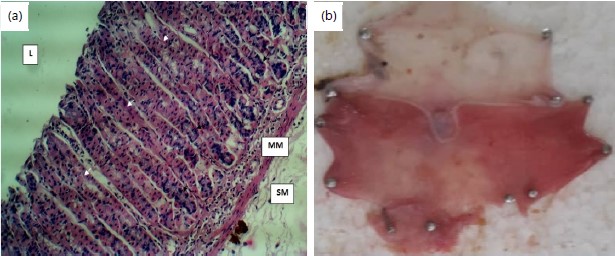
Histology: Figure 1a shows histograph section of the stomach of normal rats H&Ex160. Stomach showing normal gastric histo-architecture. Normal gastric pits, lined by the parietal cells (white arrow) and the chief cells (black arrow) were observed. Central vein (V), portal area (P), lumen (L), muscularis mucosa (MM) and submucosa (SM). Figure 1b shows a picture of stomach of group one (normal control) showing normal stomach architecture without laceration.
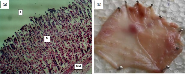
Figure 2a shows histograph section of the stomach of rats in group B (negative control) H&Ex160. Picture showed full mucosal necrosis (M). Central vein (V), portal area (P), lumen (L), muscularis mucosa (MM) and submucosa (SM). Figure 2b shows a picture of stomach of group two (negative control) showing severely lacerated stomach architecture.
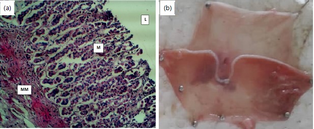
Figure 3a shows histograph section of the stomach of rats in group 3 (drug control) H&Ex160. Sections of the stomach presented in this group showed marked but improved mucosal necrosis (M). Portal area (P), lumen (L) and muscularis mucosa (MM). Figure 3b shows picture of stomach of group three (drug control) showing improved stomach architecture.
Figure 4a shows histograph section of stomach of rats in group 4 (ethanol fraction control) H&Ex160. The histograph sections of the stomach presented in this group showed slight mucosal necrosis (M). Portal area (P), lumen (L) and muscularis mucosa (MM). Figure 4b shows picture of stomach of group four (ethanol group) showing improved stomach architecture.
Figure 5a shows histograph sections of the stomach of rats in group 5 (methanol fraction) H&Ex160. The histograph sections of the stomach presented in this group showed slight mucosal necrosis (M). Portal area (P), lumen (L) and muscularis mucosa (MM). Figure 5b shows picture of stomach of group four (methanol fraction) showing improved stomach architecture.
|

|

|
|
|
|
Figure 6a shows histograph section of stomach of rats in group 6 (N-hexane fraction) H&Ex160. The histograph sections of the stomach showed normal gastric histo-architecture. Normal gastric pits, lined by the parietal cells (white arrow) and the chief cells (black arrow) were observed. Portal area (P), lumen (L), muscularis mucosa (MM) and submucosa (SM). Figure 6b shows a picture of stomach of group four showing improved stomach architecture.
DISCUSSION
The result of the study shows that the different fractions of Dialium guineense bark reduced the antioxidant activity, reversed the inflammatory cytokine concentrations and visibly reversed the gastric ulceration induced by ethanol in the study animals as confirmed by the histograph architectural display.
Gastric ulceration is a benign lesion on the mucosal epithelium upon exposure of the stomach to excess acid and aggressive pepsin activity27. Alcohol has been found to induce oxidative stress, inflammatory response and apoptosis through recruitment of leucocytes28. The NF-κB has been implicated to play a significant role in crosslinking the cascades of these events29. Alcohol has been shown to inflict hemorrhagic gastric lesions implicated by inflammatory cell infiltration, cellular exfoliation and mucosal friability, including extensive submucosal edema30. Alcohol also inflicts hemorrhage and necrotic gastric damage through mechanisms such as disruption of gastric microvessels and restriction of blood flow31.
Biochemical analysis of gastric secretions (pH) and mucosal integrity for stomach is usually employed to ascertain its status following exposure to pharmacological agents32. The pH gives an idea of the level of acidity and volume of gastric secretions. Low pH value is a manifestation of decreased hydrogen ion concentration in gastric juice33. Reduction in pH initiates negative feedback control by somatostatin hence, inhibiting gastrin release by the the antral D cells. This mechanism has been shown to play a significant role in the pathogenesis of ulcers and gastric damage in experimental animals33. Eroded mucin content is a major factor in gastrointestinal injury34. The thinning is caused by onslaughts of aggressive agents on mucosal epithelia either by internal factors (oxidants produced in the gastric lumen including pepsin) or external factors such as drugs and chemicals35.
The significant increase in ulcer index and gastric volume following oral administration of ethanol in the ulcerated rats may be attributed either to formation of free radicals or inhibition of prostaglandin synthesis16. Decreased prostaglandin level has been attributed to impaired gastroprotection and increased gastric acid secretion which are important pathways in the pathogenesis of mucosal ulceration. This agreed with the reports of Karampour et al.36 that the ulcer index of experimental animals increases after ethanol administration. The different fractions of the extract of Dialium guneense stem bark significantly reduced the index even lower than that of the drug control which indicates ulceration healing and therefore a gastroprotective effect. This healing effect could be a result of a combination of events, including release of pre-formed mucus, wound retraction and re-epithelialization which are involved in ulcer-healing process after toxicological injury33.
Since the extract caused increased synthesis and secretion of gastric mucus, it could therefore accelerate gastric ulcer healing. That was indicative of an enhanced mucus secretory potential of the extract and suggestive of its significant role in the ulcer healing process. Healing of mucosa epithelial cells was prominently displayed by all the tested fractions, thus depicting a better ulcer healing capacity when compared with the reference drug used.
Pathogenesis of diseases such as bone marrow suppression with selective megakaryocyte depression has been linked to intake of food additives37. Food additives have also been shown to induce DNA strand breakage in these cells induced by the oxidative stress leading to reductions in leucocyte and platelet counts38. The role of oxidative stress in the etiology of diseases has been reported by several studies39. Hence, the increase in reactive oxygen species generated by activated macrophages coupled with hypochlorous acid synthesized by myeloperoxidase causes ulceration of the gastric mucosa40. Tumor necrosis factor-α has been deeply implicated in gastric inflammation via many mechanisms including upregulation of NF-κB activity, activation and recruitment of immune cells and generation of other proinflammatory cytokines41. The TNF-α also suppresses gastric microcirculation around ulcerated mucosa and thus delays its healing42. The reduction of the TNF-α, PGE2 and IL-1b in the co-treated groups could be an indication of suppression of ethanol-induced toxicity. It is, therefore, reasonable to suggest that the inhibition of NF-κB is the key mechanism by which the Dialium guineense stem bark extract causes the suppression of ethanol-induced ulceration since the expression of several proinflammatory cytokines including TNF-α, PGE2 and IL-1b is mainly regulated by the transcription of NF-κB31. The NF-κB is a transcription factor that plays significant roles in toxicity, such as expression of many proinflammatory targets including adhesion molecules, TNF-α and chemokines such as PGE243. Thus Dialium guineense stem bark extract seemingly caused significant (p<0.05) protection against ethanol-induced ulceration mainly through suppression of NF-κB. This could be achieved either directly via inhibition of NF-κB target receptors such as the proinflammatory tumor necrotizing factor or indirectly through mopping up pro oxidants by the antioxidant properties of Dialium guineense stem bark extract.
Troponin is a complex of three regulatory proteins that are an integral part leading to the contraction of skeletal and cardiac muscles44. An increase in troponin has been shown to be a biomarker for onset of deep tissue injuries including pressure ulcers45. Measurements of cardiac-specific compounds are also used as diagnostic and prognostic indicators in the management of myocardial infarction and acute coronary syndrome46. Blood troponin levels may be used as a diagnostic marker for stroke or other myocardial injury that is ongoing44. The reduction in the serum troponin in the co-treated groups is an indication that Dialium guineense stem bark extract was able to ameliorate deep tissue injury (ulcer) induced by ethanol.
A dehydrogenase is an enzyme characterized by the transfer of a hydride from one molecule to another47. The LDH is expressed extensively in body tissues, such as blood cells and heart muscle48. Because it is released during tissue damage, it is a marker of common injuries and diseases, including heart failure and deep tissue injuries49. A significant increase as seen in the (negative) untreated group was an indication of oxidative challenge and agrees with the finding by of Achukwu et al.38 activity increases in conditions of deep tissue injuries. Lactate produced by anaerobic glycolysis causes a decrease in cellular pH which further exacerbates ulcer40. The significant reduction was therefore indicative that Dialium guineense stem bark extract has the potential to reduce deep tissue injuries, hence, ulcers caused by ethanol.
Creatine kinase is a known marker of early cardiac-related conditions such as cardiac arrest and myocardial infarction50. Kato et al.51 in their study, showed that creatinine kinase activity was elevated in ulcer conditions. The increase in the creatine kinase as seen in the untreated group is an indication of ulceration and agrees with the result of Tsai et al.52. However, the downregulation of the serum CK activity is an indication of an ameliorative effect and suggests that Dialium guineense stem bark extract reversed the challenge and led to the repair of the probable ulcerated stomach region.
In spite of the advances in the therapeutic management of gastric ulcers, the prevalence of this disease is still high53. Many phytochemical studies have shown that phenolic compounds possess an important role in the prevention of gastric ulcer54. Ethanol has been a common drug used in the induction of ulcers in animal models55. Ethanol intake induces gastric necrotic damage leading to inflammatory cell infiltration and reduced bicarbonate secretion. Also, ethanol inhibits gastric blood flow and increases the production of MDA and GSSG which are both implicated in oxidative stress56. It is well-known that oxidative stress and reactive oxygen species (ROS) are involved in the pathogenesis of ethanol-induced gastric ulcers. The antioxidant enzymes in all body cells consist of three major classes of antioxidant enzymes which are the catalases, (CAT), superoxide dismutases (SOD) and glutathione peroxidases (GPX), all of these, play crucial roles in maintaining homeostasis in cells and their induction reflects a specific response to pollutant oxidative stress57.
Catalase reduces oxidative stress and lipid peroxidation either by protecting the detoxifying enzymes by increasing the efficacy of Nicotineamide Dinucleotide Phosphate (NADPH) or by helping in the elimination of compounds that produce peroxidation in the cell membranes58. Since H2O2 acts as a substrate for a specific reaction that generates highly hydroxyl radicals, it is believed that the primary role of catalase in cellular antioxidant defense mechanisms is to reduce the accumulation of H2O259. The reduction in catalase activity could be a result of oxidative stress leading to the depletion of the enzyme. Thus, the increase in the enzyme activity in the co-treated groups could be attributed to the high antioxidant activity of different fractions of Dialium guineense bark extract which attenuated the ROS leading to improved antioxidant activity. Increasing the CAT activity would further deplete the ROS thereby thwarting the induction of stomach ulcers and permitting healing of the ulcers.
The role of SOD is to scavenge superoxide radicals and convert them to H2O260. Thus reduced SOD activity in the untreated group (negative control) could be an indication of oxidative stress. This agrees with the result of Nkanu et al.61 that the reduction in serum SOD activity is thought to be a result of excessive autoxidation and progressive glycation of enzymatic proteins. However, the increase in the enzyme activity in the co-treated groups could be attributed to the high antioxidant activity of the different fractions of Dialium guineense bark extract which attenuate the oxidative stress, thereby ameliorating the ethanol mechanism for ulcer induction.
The MDA is a marker for oxidative stress and a product of peroxidation of membrane lipids in plants. The result of this study indicated an increase in MDA concentration in the untreated group. This agreed with previous studies that ethanol administration in rats caused an increase in MDA concentration in animal model15. Its reduction could be indicative of gastroprotective effect of Dialium guneense bark possibly due to mopping up of free radicals, hence reducing oxidative stress. Thus, the protective effect of Dialium guneense bark on ethanol-induced gastric ulcers may be due to its potent antioxidant activity and ability to scavenge free radicals.
Glutathione-S-transferases (GST) represent a major group of antioxidant enzymes, which form a cascade of proteins implicated in the protection of tissues against oxidative damage via cellular detoxification of genotoxic and cytotoxic compounds62. Depleted GST activity is implicated in reduced activity which occurs in oxidative stress. The increase of the activity across the different fractions compared to the untreated group is attributed to an improved antioxidant state by the Dialium guineense bark methanol extract and could be a mechanism for ameliorating the ulcerative effect of ethanol.
Alteration of RBC parameters in ulcer patients are associated with different mechanisms; including decreased iron absorption secondary to chronic gastritis63, iron loss via hemorrhagic gastritis, active bleeding peptic ulcers, deficiency of iron and vitamin B12 secondary to chronic and atrophic gastritis, which might contribute to the alteration of RBC parameters64. The significant (p<0.05) lower RBC count in the negative control could be as a result of increased bleeding thus, indicative of hemorrhagic gastritis. This can be further buttressed by the histopathology examination of the stomach layer which indicated multiple lacerations. This result agreed with the works of Saler et al.65 and Haile and Timerga66, that RBC of animal models is reduced in conditions of peptic ulcer. However, the RBC count of the groups that received different fractions of the extract showed a significant (p<0.05) increase indicating that the extract ameliorated the effects either by reducing iron loss through hemorrhagic gastritis or reduced active bleeding at the sites of peptic ulcer. This result is further confirmed by histopathology examination which indicated improved physiological architecture of the stomach walls.
This study revealed a significant decrease in mean hemoglobin concentration (p<0.005) of a negative control group as compared to the control thus, agreed with previous studies67,68. Increased hepcidin production secondary to peptic ulcer decreases the release of iron from macrophages of the reticuloendothelial system and entrecote, which impairs hemoglobin synthesis69. Because hepcidin acts as an acute phase reactant in response to the inflammation produced in the gastric mucosa70. Decreased iron absorption secondary to chronic gastritis, iron loss via hemorrhagic gastritis and active bleeding peptic ulcers, are believed to contribute to a decrease of blood hemoglobin concentration and improved state shows improvements in blood hemoglobin concentration71. The increased Hb concentration in co-treated groups is indicative of improved condition and is also confirmed by the improved physiological architecture of the stomach as indicated by the histopathological examination. The five major components of the white blood cell are neutrophils, lymphocytes, monocytes, eonisophils and basophils. An increase in the WBC status is attributed to inflammation and leucopenia and is implicated in body defense challenge. An increase in WBC, PCV, Hb and RBC concentration as seen in untreated group (group 2) is suggestive of leucopenia, inflammation and general hematological abnormalities. However, the decrease in the concentrations as noticed in the groups that co-treated different fractions of Dialium guineense stem back extract could be suggestive of ameliorative potentials, hence reversal of the ulcer induced by ethanol69-71.
Measurement of enzymatic activities in the body fluids plays a pivotal role in disease diagnosis and management, determination of the extent of toxicity of a drug and could be helpful in the determination of cellular damage long before it is picked up by the conventional histological technique.
SIGNIFICANCE STATEMENT
This study discovers the evaluation of anti-ulcerogenic effects of the crude extract and fractions of Dialium guineense tree bark in ethanol-induced peptic ulcer in albino rats. This study will help the researcher uncover areas of biochemical and anti-ulcerogenic potentials of Dialium guineense tree bark in ethanol-induced peptic which many researchers were not able to explore. The result indicates that the fractions of Dialium guineense reversed stomach assault caused by ethanol administration. This will open a new chapter into research on the specific phytochemical composition of the extract which could serve as a treatment for ethanol induced ulcers. Thus a new theory on the pharmacological effects of different fractions of Dialium guineense tree bark in ethanol-induced peptic ulcer could be arrived at.
REFERENCES
- Chita, E.I and I.J. Obidike, 2020. Nutraceutical, antioxidant and hepatic histomorphological effects of Tetrapleura tetraptera leaves in monosodium glutamate-intoxicated rats. Asian J. Emerging Res., 2: 223-238.
- Zhang, K., Y. Liu, C. Wang, J. Li and L. Xiong et al., 2019. Evaluation of the gastroprotective effects of 20 (S)-ginsenoside Rg3 on gastric ulcer models in mice. J. Ginseng Res., 43: 550-561.
- Singh, A.K., S.K. Singh, P.P. Singh, A.K. Srivastava, K.D. Pandey, A. Kumar and H. Yadav, 2018. Biotechnological aspects of plants metabolites in the treatment of ulcer: A new prospective. Biotechnol. Rep., 18.
- Ismail, I.F., S. Golbabapour, P. Hassandarvish, M. Hajrezaie and N.A. Majid et al., 2012. Gastroprotective activity of Polygonum chinense aqueous leaf extract on ethanol-induced hemorrhagic mucosal lesions in rats. Evid. Based Complementary Altern. Med., 2012.
- Nsereko, G., P. Emudong, J. Omujal, J. Acai and J.M. Kungu et al., 2019. Comparison of the efficacy of crude methanolic extracts of Cassia occidentalis and Euphorbia hirta with levamisole-HCL against gastrointestinal nematodes of economic importance to goat production in Uganda. Trop. Anim. Health Prod., 51: 2269-2278.
- Gang, C., H. Yueqing, F. Yuan, W. Aiping and L. Xuezheng et al., 2019. Extract of Ilex rotunda Thunb alleviates experimental colitis-associated cancer via suppressing inflammation-induced miR-31-5p/YAP overexpression. Phytomedicine, 62.
- Agbaje, E.O. and Y.P. Doe, 2015. Gastric and duodenal antiulcer effects of aqueous bark extract of Dialium guineense Wild. (Fabaceae) and the possible mechanisms in laboratory models. J. Phytopharmacol., 4: 268-275.
- Ezeja, M.I., Y.S. Omeh, I.I. Ezeigbo and A. Ekechukwu, 2011. Evaluation of the analgesic activity of the methanolic stem bark extract of Dialium guineense (Wild). Ann. Med. Health Sci. Res., 1: 55-62.
- Besong, E.E., M.E. Balogun, S.F.A. Djobissie, D.C. Obu and J.N. Obimma, 2016. Medicinal and economic value of Dialium guineense. Afr. J. Biomed. Res., 19: 163-170.
- Assiki, T., A. Diallo, E. Badjabaïssi, M. Assih and T. Kpatcha et al., 2022. Antidiarrheal activity of Dialium guineense willd fruit pulp in Wistar rats. BioMed Res. Int., 2022.
- Ayoub, F., V. Khullar, D. Banerjee, P. Stoner and T. Lambrou et al., 2018. Once versus twice-daily oral proton pump inhibitor therapy for prevention of peptic ulcer rebleeding: A propensity score-matched analysis. Gastroenterol. Res., 11: 200-206.
- Ray-Offor, E. and K.A. Opusunju, 2020. Current status of peptic ulcer disease in Port Harcourt metropolis, Nigeria. Afr. Health Sci., 20: 1446-1451.
- Irabor, D.O., 2005. An audit of peptic ulcer surgery in Ibadan, Nigeria. West Afr. J. Med., 24: 242-245.
- Narayanan, M., K.M. Reddy and E. Marsicano, 2018. Peptic ulcer disease and Helicobacter pylori infection. Mo. Med., 115: 219-224.
- Shams, S.G.E. and R.G. Eissa, 2022. Amelioration of ethanol-induced gastric ulcer in rats by quercetin: Implication of Nrf2/HO1 and HMGB1/TLR4/NF-κB pathways. Heliyon, 8.
- Mousa, A.M., N.M. El-Sammad, S.K. Hassan, A.E.N.A. Madboli and A.N. Hashim et al., 2019. Antiulcerogenic effect of Cuphea ignea extract against ethanol-induced gastric ulcer in rats. BMC Complementary Altern. Med., 19.
- Bhattacharyya, A., R. Chattopadhyay, S. Mitra and S.E. Crowe, 2014. Oxidative stress: An essential factor in the pathogenesis of gastrointestinal mucosal diseases. Physiol. Rev., 94: 329-354.
- Tarnawski, A.S. and A. Ahluwalia, 2021. The critical role of growth factors in gastric ulcer healing: The cellular and molecular mechanisms and potential clinical implications. Cells, 10.
- Yanaka, A., 2018. Role of NRF2 in protection of the gastrointestinal tract against oxidative stress. J. Clin. Biochem. Nutr., 63: 18-25.
- Liu, C.M., J.Q. Ma, W.R. Xie, S.S. Liu, Z.J. Feng, G.H. Zheng and A.M. Wang, 2015. Quercetin protects mouse liver against nickel-induced DNA methylation and inflammation associated with the Nrf2/HO-1 and p38/STAT1/NF-κB pathway. Food Chem. Toxicol., 82: 19-26.
- Aziz, R.S., A. Siddiqua, M. Shahzad, A. Shabbir and N. Naseem, 2019. Oxyresveratrol ameliorates ethanol-induced gastric ulcer via downregulation of IL-6, TNF-α, NF-ĸB, and COX-2 levels, and upregulation of TFF-2 levels. Biomed. Pharmacother., 110: 554-560.
- Johnlouis, O.I., N.C. Ifeanyi and O.P. Oluchukwu, 2022. Consequence of Tetrapleura tetraptera leaves on pro-oxidants, hepatic functions and histomorphology in monosodium glutamate-intoxicated rats. Res. J. Med. Plants, 16: 37-48.
- Nwafor, P.A., F.K. Okwuasaba and L.G. Binda, 2000. Antidiarrhoeal and antiulcerogenic effects of methanolic extract of Asparagus pubescens root in rats. J. Ethnopharmacol., 72: 421-427.
- Budimir, I., S. Stojsavljević, N. Baršić, A. Bišćanin and G. Mirošević et al., 2017. Scoring systems for peptic ulcer bleeding: Which one to use? World J. Gastroenterol., 23: 7450-7458.
- Obidike, I.J. and M.E. Ngozi, 2024. Effects of carrot (Daucus carota) stalk on blood glucose level, biochemical functions and serum antioxidant activity in alloxan-induced diabetic rats. Asian Sci. Bull., 2: 46-59.
- Johnlouis, O.I., C.E. ije, N.C. Ifeanyi and O.Q. Chiamaka, 2023. Effects of Tetrapleura tetraptera leaves on renal architecture and haematological indices in monosodium glutamate-intoxicated rats. Asian Sci. Bull., 1: 8-17.
- Khazaei, M. and H. Salehi, 2006. Protective effect of Falcaria vulgaris extract on ethanol induced gastric ulcer in rat. Iran. J. Pharmacol. Ther., 5: 43-46.
- Simões, S., R. Lopes, M.C.D. Campos, M.J. Marruz, M.E.M. da Cruz and L. Corvo, 2019. Animal models of acute gastric mucosal injury: Macroscopic and microscopic evaluation. Anim. Models Exp. Med., 2: 121-126.
- Sangiovanni, E., U. Vrhovsek, G. Rossoni, E. Colombo and C. Brunelli et al., 2013. Ellagitannins form Rubus berries for the control of gastric inflammation: In vitro and in vivo studies. PLoS ONE, 8.
- Park, S.W., T.Y. Oh, Y.S. Kim, H. Sim and S.J. Park et al., 2008. Artemisia asiatica extracts protect against ethanol-induced injury in gastric mucosa of rats. J. Gastroenterol. Hepatol., 23: 976-984.
- Li, W., H. Huang, X. Niu, T. Fan, Q. Mu and H. Li, 2013. Protective effect of tetrahydrocoptisine against ethanol-induced gastric ulcer in mice. Toxicol. Appl. Pharmacol., 272: 21-29.
- Adhikary, B., S.K. Yadav, K. Roy, S.K. Bandyopadhyay and S. Chattopadhyay, 2011. Black tea and theaflavins assist healing of indomethacin-induced gastric ulceration in mice by antioxidative action. Evidence-Based Complementary Altern. Med., 2011.
- Sabiu, S., T. Garuba, T. Sunmonu, E. Ajani, A. Sulyman, I. Nurain and A. Balogun, 2015. Indomethacin-induced gastric ulceration in rats: Protective roles of Spondias mombin and Ficus exasperate. Toxicol. Rep., 2: 261-267.
- Inas, Z.A.A., A.H.K. Hala, and H.H. Gehan, 2011. Gastroprotective effect of assyrian plum (Cordia myxa L.) fruit extract against indomethacin-induced gastric ulceration in rats. Life Sci. J., 8: 433-445.
- Zhou, D., Q. Yang, T. Tian, Y. Chang and Y. Li et al., 2020. Gastroprotective effect of gallic acid against ethanol-induced gastric ulcer in rats: Involvement of the Nrf2/HO-1 signaling and anti-apoptosis role. Biomed. Pharmacother., 126.
- Karampour, N.S., A. Arzi, A. Rezaie, M. Pashmforoosh and F. Kordi, 2019. Gastroprotective effect of zingerone on ethanol-induced gastric ulcers in rats. Medicina, 55.
- Gutiérrez, O.M., A. Luzuriaga-McPherson, Y. Lin, L.C. Gilbert, S.W. Ha and G.R. Beck, 2015. Impact of phosphorus-based food additives on bone and mineral metabolism. J. Clin. Endocrinol. Metab., 100: 4264-4271.
- Achukwu, P.U., S.A. Ufelle, E.O. Ukaejiofo, F.E. Ejezie and D.N. Nwachukwu et al., 2009. The Effect of Potassium Bromate on Some Haematological Parameters of Wistar Rats. Niger. J. Physiol. Sci., 24: 59-61.
- Mansour, D.F., D.O. Saleh, O.A. Ahmed-Farid, M. Rady, R.M. Bakeer and I.M. Hashad, 2021. Ginkgo biloba extract (EGb 761) mitigates methotrexate-induced testicular insult in rats: Targeting oxidative stress, energy deficit and spermatogenesis. Biomed. Pharmacother., 143.
- Li, G., J. Gao, Y.L. Tao, B.Q. Xu and Z.W. Tu et al., 2012. Increased pretreatment levels of serum LDH and ALP as poor prognostic factors for nasopharyngeal carcinoma. Chin. J Cancer, 31: 197-206.
- Yadav, S.K., B. Adhikary, S. Chand, B. Maity, S.K. Bandyopadhyay and S. Chattopadhyay, 2012. Molecular mechanism of indomethacin-induced gastropathy. Free Radical Biol. Med., 52: 1175-1187.
- Hasgul, R., S. Uysal, H. Haltas, S. Akyol, Y. Yuksel, A. Gurel and F. Armutcu, 2014. Protective effects of Ankaferd blood stopper on aspirin-induced oxidative mucosal damage in a rat model of gastric injury. Toxicol. Ind. Health, 30: 888-895.
- Mei, X., D. Xu, S. Xu, Y. Zheng and S. Xu, 2012. Novel role of Zn (II)-curcumin in enhancing cell proliferation and adjusting proinflammatory cytokine-mediated oxidative damage of ethanol-induced acute gastric ulcers. Chem. Biol. Interact., 197: 31-39.
- Roos, A., U. Sartipy, R. Ljung and M.J. Holzmann, 2018. Relation of chronic myocardial injury and non-ST-segment elevation myocardial infarction to mortality. Am. J. Cardiol., 122: 1989-1995.
- Traa, W.A., G.J. Strijkers, D.L. Bader and C.W.J. Oomens, 2019. Myoglobin and troponin concentrations are increased in early stage deep tissue injury. J. Mech. Behav. Biomed. Mater., 92: 50-57.
- Strandberg, L.S., A. Roos and M.J. Holzmann, 2021. Stable high-sensitivity cardiac troponin T levels and the association with frailty and prognosis in patients with chest pain. Am. J. Med. Open, 1-6.
- Stacpoole, P.W. and C.E. McCall, 2023. The pyruvate dehydrogenase complex: Life’s essential, vulnerable and druggable energy homeostat. Mitochondrion, 70: 59-102.
- Elsea, S.H., J. Razjouyan, K.M. Lee, J.A. Lynch, S. Martini and L.M. Pandit, 2023. Association of glucose-6-phosphate dehydrogenase deficiency with outcomes in us veterans with COVID-19. JAMA Netw. Open, 6.
- Zhang, Y., K. Chen, S.A. Sloan, M.L. Bennett and A.R. Scholze et al., 2014. An RNA-sequencing transcriptome and splicing database of glia, neurons, and vascular cells of the cerebral cortex. J. Neurosci., 34: 11929-11947.
- Kristjansson, R.P., A. Oddsson, H. Helgason, G. Sveinbjornsson and G.A. Arnadottir et al., 2016. Common and rare variants associating with serum levels of creatine kinase and lactate dehydrogenase. Nat. Commun., 7.
- Kato, A., H. Naitou, M. Namioka, M. Akimoto and T. Ishii et al., 2010. Proteomic identification of serum proteins associated with stress-induced gastric ulcers in fasted rats. Biosci. Biotechnol. Biochem., 74: 812-818.
- Tsai, S.H., S.J. Chu, C.W. Hsu, S.M. Cheng and S.P. Yang, 2008. Use and interpretation of cardiac troponins in the ED. Am. J. Emergency Med., 26: 331-341.
- Rodríguez, J.A., C. Theoduloz, T. Yáñez, J. Becerra and G. Schmeda-Hirschmann, 2006. Gastroprotective and ulcer healing effect of ferruginol in mice and rats: Assessment of its mechanism of action using in vitro models. Life Sci., 78: 2503-2509.
- Sumbul, S., M.A. Ahmad, M. Asif and M. Akhtar, 2011. Role of phenolic compounds in peptic ulcer: An overview. J. Pharm. BioAllied Sci., 3: 361-367.
- Arab, H.H., S.A. Salama, H.A. Omar, E.S.A. Arafa and I.A. Maghrabi, 2015. Diosmin protects against ethanol-induced gastric injury in rats: Novel anti-ulcer actions. Plos One, 10.
- El-Maraghy, S.A., S.M. Rizk and N.N. Shahin, 2015. Gastroprotective effect of crocin in ethanol-induced gastric injury in rats. Chem. Biol. Interact., 229: 26-35.
- Birben, E., U.M. Sahiner, C. Sackesen, S. Erzurum and O. Kalayci, 2012. Oxidative stress and antioxidant defense. World Allergy Organ. J., 5: 9-19.
- Abd-Elkareem, M., M. Soliman, M.A.M. Abd El-Rahman and N.S. Abou Khalil, 2022. The protective effect of Nigella sativa seeds against monosodium glutamate-induced hepatic dysfunction in rats. Toxicol. Rep., 9: 147-153.
- Htet, A.S., M.K. Kjøllesdal, W.P. Aung, A.N.M. Myint and W.T. Aye et al., 2017. Lipid profiles and determinants of total cholesterol and hypercholesterolaemia among 25-74 year-old urban and rural citizens of the Yangon Region, Myanmar: A cross-sectional study. BMJ Open, 7.
- Hayyan, M., M.A. Hashim and I.M. AlNashef, 2016. Superoxide ion: Generation and chemical implications. Chem. Rev., 116: 3029-3085.
- Nkanu, E.E., G. Ujong, V. Okon and I.E. Bassey, 2018. In vivo efficacy of Dialium guineense fruit pulp on hemeoxygenase-1 and angiotensin converting enzyme in experimental diabetes. J. Med. Plants Res., 12: 483-492.
- Muid, K.A., H.C. Karakaya and A. Koc, 2014. Absence of superoxide dismutase activity causes nuclear DNA fragmentation during the aging process. Biochem. Biophys. Res. Commun., 444: 260-263.
- Al Mutawa, O.A., M.A. Izhari, R.A. Alharbi, A.A.A. Sindi and A.M. Alqarni et al., 2023. Helicobacter pylori (H. pylori) infection-associated anemia in the Asir Region, Saudi Arabia. Diagnostics, 13.
- Mărginean, C.D., C.O. Mărginean and L.E. Meliț, 2022. Helicobacter pylori-related extraintestinal manifestations-myth or reality. Children, 9.
- Saler, T., Ş.Ö. Keşkek, S. Kırk, S. Ahbab and G. Ortoğlu, 2014. H. pylori may not be associated with iron deficiency anemia in patients with normal gastrointestinal tract endoscopy results. Adv. Hematol., 2014.
- Haile, K. and A. Timerga, 2021. Evaluation of hematological parameters of Helicobacter pylori-infected adult patients at Southern Ethiopia: A comparative cross-sectional study. J. Blood Med., 12: 77-84.
- Mwafy, S.N. and W.M. Afana 2018. Hematological parameters, serum iron and vitamin B12 levels in hospitalized Palestinian adult patients infected with Helicobacter pylori: A case-control study. Hematol. Transfus. Cell Ther. 40: 160-165.
- Barroso, C., P. Carvalho, M. Nunes, J.F.M. Gonçalves, P.N.S. Rodrigues and J.V. Neves, 2021. The era of antimicrobial peptides: Use of hepcidins to prevent or treat bacterial infections and iron disorders. Front. Immunol., 12.
- Santambrogio, E. and L. Orsucci, 2019. Helicobacter pylori and hematological disorders. Minerva Gastroenterol. Dietologica, 65: 204-213.
- Nasif, W.A., A.S.E. Ali, H.S. Alamodi, A.A. Alrefai and A.A. Alzubedi et al., 2021. Impact of Helicobacter pylori on hematological parameters among Saudi population. Saudi Med. J., 42: 643-648.
- Moldovan, O.L., C.E. Vari, A. Tero-Vescan, O.S. Cotoi and I.G. Cocuz et al., 2023. Potential defence mechanisms triggered by monosodium glutamate sub-chronic consumption in two-year-old Wistar rats. Nutrients, 15.
How to Cite this paper?
APA-7 Style
Johnlouis,
O.I., Sunday,
A.G., Adanma,
O.C., Ngozi,
O.J. (2024). Evaluation of Anti-Ulcerogenic Effects of the Crude Extract and Fractions of Dialium guineense Tree Bark in Ethanol-Induced Peptic Ulcer in Albino Rats. Asian Journal of Biological Sciences, 17(3), 254-273. https://doi.org/10.3923/ajbs.2024.254.273
ACS Style
Johnlouis,
O.I.; Sunday,
A.G.; Adanma,
O.C.; Ngozi,
O.J. Evaluation of Anti-Ulcerogenic Effects of the Crude Extract and Fractions of Dialium guineense Tree Bark in Ethanol-Induced Peptic Ulcer in Albino Rats. Asian J. Biol. Sci 2024, 17, 254-273. https://doi.org/10.3923/ajbs.2024.254.273
AMA Style
Johnlouis
OI, Sunday
AG, Adanma
OC, Ngozi
OJ. Evaluation of Anti-Ulcerogenic Effects of the Crude Extract and Fractions of Dialium guineense Tree Bark in Ethanol-Induced Peptic Ulcer in Albino Rats. Asian Journal of Biological Sciences. 2024; 17(3): 254-273. https://doi.org/10.3923/ajbs.2024.254.273
Chicago/Turabian Style
Johnlouis, Obidike, Ikechukwu, Aloh Godwin Sunday, Obike Chiemeziem Adanma, and Okechukwu Jane Ngozi.
2024. "Evaluation of Anti-Ulcerogenic Effects of the Crude Extract and Fractions of Dialium guineense Tree Bark in Ethanol-Induced Peptic Ulcer in Albino Rats" Asian Journal of Biological Sciences 17, no. 3: 254-273. https://doi.org/10.3923/ajbs.2024.254.273

This work is licensed under a Creative Commons Attribution 4.0 International License.