Phytochemical Profiling, Antioxidant and Anti-Cancerous Activity of Hydrogonium arcuatum (Griff.) Wijk. & Marg. (Bryophyta: Pottiaceae)
-
Tripti Sharma
Department of Bioscience and Biotechnology, Banasthali Vidyapith (Rajasthan), India
Swati SinghDepartment of Bioscience and Biotechnology, Banasthali Vidyapith (Rajasthan), India
Afroz Alam

Department of Bioscience and Biotechnology, Banasthali Vidyapith (Rajasthan), India
| Received 04 Apr, 2024 |
Accepted 11 Sep, 2024 |
Published 30 Sep, 2024 |
Background and Objective: Bryophytes, the second-largest category of plants make up a considerable portion of biodiversity found in arid regions. This investigation aims to reveal the antioxidant capacity and phytochemical makeup of Hydrogonium arcuatum thereby enhancing understanding of its biological attributes. Materials and Methods: After the gametophytic thalli were removed from their natural habitats and thoroughly cleaned, extracts were made using normal procedures. Using well-established protocols linked to GC-MS, TLC and FTIR techniques, phytochemical profiling, antioxidant activity and anti-cancerous activities were evaluated and results were observed. Results: Methanolic extract contained tannins, alkaloids, flavonoids and phenols, according to phytochemical screening. In contrast, a confirmatory test using the TLC method revealed the presence of flavonoids and alkaloids. Methanolic extract yielded the greatest results in the antioxidant experiment, followed by ethyl acetate extract. Caryophyllene, which has been demonstrated to have antiviral, anti-inflammatory and cytotoxic qualities, is confirmed to be present by the GC-MS approach. At 1000 µg/mL, H. arcuatum extract showed the greatest cytotoxicity against HT-29, but not against HCT-116. At dosages of 50 and 100 µg/mL, it did not affect either of the two cell lines. Conclusion: The H. arcuatum has been shown to exhibit a highly significant increase in cytotoxicity against cell lines, because of its strong cytotoxic action. This suggests that it may be a valuable treatment option for colorectal cancer.
| Copyright © 2024 Sharma et al. This is an open-access article distributed under the Creative Commons Attribution License, which permits unrestricted use, distribution, and reproduction in any medium, provided the original work is properly cited. |
INTRODUCTION
Bryophytes, the second-largest category of plants after angiosperms, make up a considerable portion of biodiversity, with 16,600 species worldwide and 850 species in India1,2. These plants are classified into three main groups: Hornworts (Anthocerotopsida), liverworts (Marchantiopsida) and mosses (Bryopsida). They are phylogenetically located between algae and pteridophytes3-5.
Bryophytes are distributed around the world in a variety of biological environments, from the tropics to the polar and alpine regions. In our country, they are primarily found in the Eastern and Western
Himalayas, South India and Central India6,7. They are divided into 21 orders that benlong to 66 families, which contain 328 genera and 1578 species8-10.
They are used in the manufacture of furniture and household items, gardening, the pharmaceutical industry (e.g., for surgical dressings, medications, antibiotics, etc.) and as packing material11,12. The second-most specious category after angiosperms, mosses make up a sizeable portion of the diversity (ca. 13,000 species) of terrestrial plants13. They can even be found in many harsh settings where vascular plants are few or nonexistent.
Since it is well known that, in contrast to higher vascular plants, bryophytes lack any type of mechanical or physical protection, they have an excellent metabolism and remarkable defense mechanisms to withstand a variety of biotic and abiotic challenges. Due to this, the majority of bryophytes create a variety of secondary metabolites with distinctive flavors or aromas; these secondary metabolites also have antibacterial properties that are effective against bacteria and fungi14.
Phytochemicals are plant-derived secondary metabolites. Several bryophytes have been examined for the existence of diverse phytochemicals including flavonoids, organic acids, sterols, phenolic acids, triterpenes, lipids and aldehydes. These substances are created synthetically and stored in very high concentrations12,15. Many terpenoids with about 1400 structures have been isolated from bryophytes as secondary metabolites, viz., pinguisanes, myltaylanes and ventricosanes16-19. Apart from this, a lot of different types of secondary metabolites seemed to be isolated from bryophytes. These secondary metabolites have been analyzed for their respective functions. All terpenoid classes, such as monoterpenoids, diterpenoids, sesquiterpenoids and etheric oils, are isolated from mosses and liverworts.
Among all secondary metabolites, flavonoids are considered the most interesting polyphenols in the defense system of bryophytes, as they help to combat various environmental stresses20. Bryophytes are a good source of tetraterpenoid carotenoids; many of them have been isolated from species of mosses and liverwort. Previously, it was investigated that bryophytes possess an excessive amount of secondary metabolites such as terpenoids, phenolics (flavonoids and bibenzyle derivatives), glycosides, fatty acids, as well as some aromatic compounds21,22. Whereas in few tropical moss plants, the presence of alkaloids, flavonoids, phenols, saponins and steroids was investigated, which could be a possible source of beneficial drugs in the treatment of chronic diseases23.
In the human body, the production of reactive oxygen species (ROS), superoxide and hydrogen peroxide is the most common physiological process24. A high level of ROS can be produced by exogenous chemicals and endogenous metabolic processes, which are directly connected to cardiovascular diseases such as hypertension25, diabetes and atherosclerosis26. Cirrhosis, emphysema, genotoxicity, inflammation and cancer have also been correlated with ROS effects27. Presently, it is widely acknowledged that many plant extracts and phytochemicals have specific antibacterial properties and can be useful in therapeutic treatments9. In order to show its effectiveness, many studies have been carried out recently in many countries, including India28. Mosses have shown antioxidant properties which were assayed by the DPPH method29.
Since mosses have the potential to be a source of a wide range of secondary metabolites, they are successfully incorporated into a range of pharmaceutical goods, such as antibiotics, surgical dressings and herbal treatments. Studies based on the phytochemistry of bryophytes show that a variety of biologically active substances, including organic acids, aliphatic compounds, carbohydrates, proteins, steroids, lipids, terpenoids, polyphenols, fatty acids, sugar alcohol, acetogenins, phenyl-quinines, phenolics and aromatic
compounds, are present in bryophytes and are responsible for their many bioactivities15,30. In India, burned ash of mosses is mixed with honey and used as an ointment for cuts, wounds and burns12. These pharmacological effects include antimicrobial, antifungal, cytotoxic, antitumor, vasopressin antagonist, cardiotonic, allergy-causing, insecticidal and piscicidal22.
The main objective of this study was to assess the phytochemical constituents of the moss, Hydrogonium arcuatum from Mount Abu, Rajasthan. Qualitative assays determined the presence of phenol, flavonoid and alkaloids in the methanolic extracts of H. arcuatum and then subsequent quantitative assays were performed to determine the total phenolic content (TPC), total flavonoid content (TFC) and total alkaloid content (TAC) in the methanolic extracts. Further GC- MS and TLC analyses were performed to ascertain active compounds in these extracts. The DPPH assay and nitric oxide scavenging activity assay were performed for its anti- oxidant potential. The functional groups of phytoconstituents found in the plant extract were determined by FTIR assay. Cytotoxic effect of the methanolic extract of H. arcuatum was studied against two human colorectal cancer cell lines HT-29 and HCT-116.
MATERIALS AND METHODS
Collection of plant: The plant material, i.e., selected moss species of the family Pottiaceae, was collected from Mount Abu, western Rajasthan, at an altitude of 1600 m, 24°31‘ to 24°43‘N and 72°38‘ to 72°53‘E; during the months of August 2022, subsequently in September 2022 some samples were procured from the Banasthali University Rajasthan, India (BURI) Herbarium. The collected materials were kept in brown paper packets directly in the case of dried specimens and for wet specimens, blotting paper was used to soak up excess moisture and dry the specimens. For the morpho-taxonomic study, the collected plant specimens were air-dried at room temperature and kept in brown paper herbarium packets (size, “6×4’’ inches). The morpho-taxonomic work was completed in the first week of October 2022, followed by the initiation of phytochemical work in the second week of October, which was completed in November 2022.
Identification and screening of plant material: In order to identify the samples, morphological study of plants was done in October 2022 using the Olympus Japan SZ-PT Stereoscopic Zoom Binocular Microscope to examine the plants' external morphology and the Olympus OIC HC 10068 Compound Microscope and Olympus CH 20i Binocular Compound Microscope were used to examine the cellular details and microscopic structures (No. 76026946 for the Nikon Coolpix L21). Identifications were made following careful examination of the specimens, with the assistance of the insightful contributions of former bryologists, current literature and herbarium deposited with pertinent data in the Banasthali University, Rajasthan, India (BURI) herbarium (BURI-1717/2023).
The samples were first stretched by soaking them in lukewarm water for at least half an hour at room temperature before being used to study. Samples were studied microscopically for gametophytes like leaves, stems and rhizoids. The identification of these species was done with the help of previously available specimens and literature.
Preparation of plant extracts: For the preparation of plant extract, standard procedures were followed31. Prior to extraction, all the identified samples were thoroughly washed with running tap water to free them from any adhering soil and debris and then they were finally rinsed with distilled water and air-dried in the shade until the water content became almost nil. Washed and air-dried plant samples were further ground into a fine powder with the help of a mortar and pestle using solvents (methanol, ethyl-acetate, di-ethyl ether and n- hexane) and subjected to 48 hrs of 37°C shaking at 120 rpm in an orbital shaker, the Metrex-100C. After centrifuging each extract for 30 minutes at 10,000 rpm, the supernatant was collected and stored at 4°C for further use32.
Phytochemical assay
Preliminary qualitative analysis: Using established techniques, the preliminary phytochemical analysis of methanol, ethyl acetate, diethyl ether and hexane was checked for the presence of several phytochemicals in a number of bryophytes33,34.
Quantitative analysis
Estimation of total phenolic content (TPC): Total phenolics were quantified colorimetrically using the Folin-Ciocalteu method35. The 0.5 mL of water and 0.125 mL of the methanolic extract were put into a test tube. In that sequence, 0.125 mL of the Folin-Ciocalteu reagent, 1.25 mL of sodium carbonate solution and 3 mL of water were added. The mixture was then allowed to stand for 90 min. The absorbance was recorded at 760 nm. Gallic acid equivalents (GAE) based on dry material were used to calculate the total phenol concentration (mg GAE/g dry weight of sample). These tests were conducted in triplicate.
Estimation of total flavonoid content (TFC): The approach of Adebiyi et al.23 (2016) was used to determine the total flavonoid content. The plant extract was mixed with 10% aluminum chloride, 95% ethanol, 1M potassium acetate and distilled water. The reaction mixture was allowed to stand at room temperature for thirty minutes. At 415 nm, the absorbance was recorded spectrophotometrically. The extracts' dry weight in milligrams of quercetin equivalent (mgQE/g) was used to express the results.
Estimation of total alkaloid content (TAC): The total alkaloid content (TAC) was calculated using a spectrophotometric method36. This process relates to the reaction between alkaloids and bromocresol green (BCG). The plant sample was filtered after being individually dissolved in 1 mg/mL and 2 N-HCl solvents. The pH of the phosphate buffer mixture was neutralized with 0.1 N NaOH. As 5 mL of BCG (bacillus Calmette-Guérin) solution and 5 mL of phosphate buffer were each added to 1 mL of this mixture in a separating funnel. Chloroform was used to extract the resulting complex after the mixture had been vigorously stirred. Chloroform was further used to dilute the sample to volume in a 10 mL volumetric flask. The complex's chloroform absorbance was recorded at 470 nm.
Gas Chromatography-Mass Spectroscopy (GC–MS) analysis: The GC-MS analysis was performed as per the protocol described by Abu Bakar et al.37. A Thermo Scientific Triple Quadruple (trace 1300, Tsq 8000 triple quadruple MS) GC-MS instrument was used for the analysis. Initially, the column temperature was set to 50°C for 4 min followed by an increase to 320°C for 20 min at a rate of 7°C per min. The sample injection volume was kept at 0.11, split mode was 20:1 and the temperature of the injector was then set at 280°C. Helium was used as carrier gas and the flow rate was maintained at 1 mL/min and the run time was 60 min. The method of obtaining mass spectra in the m/z 40-700 range involved electron ionization at a potential of 70 eV. The sample's chromatogram was identified by comparing the mass spectra to the NIST (National Institute of Standards and Technology) library data and the GC retention time to the recognized standards.
Thin layer chromatography (TLC) analysis of secondary metabolites: On the basis of the findings of the qualitative phytochemical analysis, TLC of the samples was performed using known standards. A precisely measured volume of extract was dissolved in methanol solvent to obtain a known concentration. The standard silica gel 60F254 aluminum sheet (310 cm–1) was used to separate the extract into an appropriate mobile phase. A microcapillary tube was used to spot the sample on the aluminum sheet. Various combinations of solvent systems were used to separate the compounds and their varied polarity was examined as a result. With a solvent system that contained an ethyl acetate: methanol volume ratio of (85:15), a chromatogram was created. The TLC sheet was lowered into the chosen solvent system's chamber and the solvent was allowed to soak through to the bottom third of the sheet. It was then removed from the chamber and air-dried. To see the spots, the TLC sheet was then kept in the UV room.
After that, a particular kind of compound was finished, with one compound erectly separated spots being the result. The amount of space shifted over the entire amount of space covered by the solvent is comparable to the retention factor (Rf) for each point38.
Antioxidant assay
DPPH (2, 2-Diphenyl-1-Picrylhydrazy) radical scavenging activity: The procedure outlined by Pejin et al.39 was utilized to ascertain the impact of the crude methanolic extract on free radicals. As 4 g of DPPH, were dissolved in 100 mL of methanol. The extract was combined with 2 mL of DPPH solution. Using an ELICO double beam SL 210 UV Vis Spectrophotometer at 517 nm, the decrease in the DPPH free radical was assessed after the 30 min incubation period. The IC50 value was used to calculate the scavenging capacity of the methanolic extract. It is described as a sample concentration that causes a 50% decrease in oxidative radicals. The strength of the antioxidant scavenging activity increases with decreasing IC50 values. The extract's scavenging activity was evaluated using the percentage of decolorization. The sample's scavenging activity was calculated using the sample's percentage of decolorization.
Nitric oxide scavenging activity: The 2 mL Sodium nitroprusside (10 mM) in 0.5 mL phosphate buffer saline (1M; pH 7.4) was mixed with 0.5 mL extract and incubated for 150 min at 25°C. The 0.5 mL of the incubated mixture was taken and mixed with 1.0 mL of sulfanilic acid reagent. Finally, 1.0 mL of 0.1% naphthylethylenediamine dihydrochloride was mixed and incubated for 30 min at room temperature. The absorbance was recorded at λ540 nm. The NOSA was calculated and expressed as IC50 (μg/mL) from the method of Badami et al.40
Fourier Transform Infrared Spectrophotometer analysis (FTIR): Four species of moss extracts were directly loaded into the FTIR spectrum and a spectrometer was obtained from Shamran et al.41 The FTIR spectrometer (Thermo Scientific) had a wavelength range of 400 to 4000 cm–1 with a resolution of 4 cm–1 and this wavelength range was used to determine the functional groups of phytoconstituents found in the plant extract.
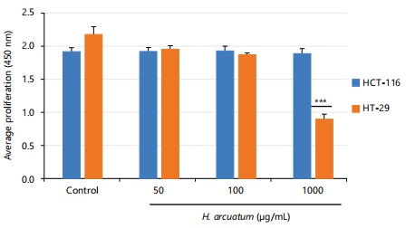
Assessment of cytotoxicity: For the cytotoxicity assessment, two human colorectal cancer cell lines namely, HT-29 and HCT-116 were procured from ATCC, Manassas, VA. Routine propagation of cell lines was done as per the standard protocol42. Cell viability was assessed by the XTT assay. Absorbance was recorded by a microplate reader at λ660 nm and λ475 nm.
Statistical analysis: The mean (n = 3) of three replicates has been used to present the data. The collected data was analyzed using IBM SPSS Statistical 20 software. The data were compared using one-way ANOVA followed by the Tukey’s test, with a significance level of less than 0.05. The data presented is expressed as a standard deviation.
RESULTS
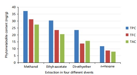
Phytochemical screening: In qualitative analysis alkaloids, phenol, flavonoid, tannins and terpenoids were present in the methanolic extract of H. arcuatum. Gallic acid, quercetin and atropine were used to express the total content of phenol, flavonoid and alkaloid of H. arcuatum (Table 1-2; Fig. 1). Large amounts of phenol, flavonoid and alkaloids were produced from H. arcuatum using methanol as a solvent; however, much lower amounts of these contents were produced using n-hexane as a solvent.
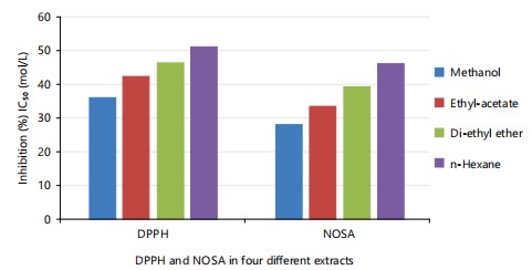
Antioxidant assay: The antioxidant activity of the plant extract as IC50 value was determined to be high potential in methanol followed by ethyl acetate (μg/mL; Mean±SD, n = 3) against DPPH and NOSA, respectively (Table 3; Fig. 2).
|
|
| Table 1: | Qualitative analysis of Hydrogonium arcuatum in different extracts | |||
| Presence and absence of Hydrogonium arcuatum in different extracts | |||||
| Phytochemicals | Test | n- hexane | Diethyl ether | Ethyl acetate | Methanol |
| Alkaloids | Dragendorff’s reagent test | - | + | ++ | +++ |
| Phenols | Ferric chloride test | + | + | ++ | +++ |
| Saponin glycosides | Froth formation test | - | - | - | - |
| Cardiac glycosides | Kellar- Killani test | - | - | - | - |
| Tannins | Ferric chloride test | - | + | ++ | +++ |
| Proteins | Xanthoprotein test | - | - | + | +++ |
| Steroids | Salkowski test | - | - | - | - |
| Terpenoids | Salkowski test | + | + | ++ | +++ |
| Amino acids | Millon’s test | - | + | + | +++ |
| Carbohydrate | Molisch’s test | - | + | ++ | +++ |
| Fats | Saponification test | - | - | + | ++ |
| Flavonoids | Shinoda test | + | + | ++ | +++ |
| Anthraquinone | Borntrager’s test | - | - | + | ++ |
| +++: High presence, ++: Moderate presence, +: Low presence and -: Absent | |||||
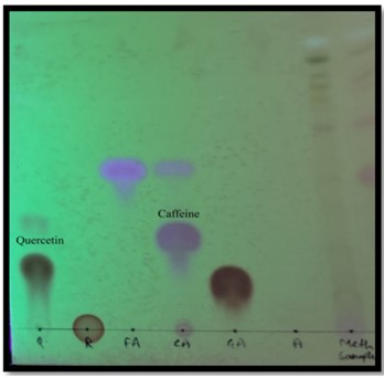
Thin layer chromatography: Thin layer chromatography was used to separate different chemicals from a methanolic extract of H. arcuatum and the findings show four spots with distinct colors and Rf values calculated by comparing the solute and solvent travel distances. The spots that appeared on sheet were calculated with Rf values of 0.42 and 0.53, respectively, which confirms the existence of quercetin and caffeine as per standard values (Table 4; Fig. 3).
|
| Table 2: | Quantitative analysis of Hydrogonium arcuatum extract in four solvents | |||
| Solvents variable | Methanol | Ethyl-acetate | Di-ethyl ether | n-Hexane |
| TPC | 38.05±0.29c mg/GAE/g | 31.02±0.17b mg/GAE/g | 24.02±0.11b mg/GAE/g | 12.04±0.04a mg/GAE/g |
| TFC | 32.02 ±0.23c mg/QE/g | 24.06 ±0.19c mg/QE/g | 14.04 ±0.08b mg/QE/g | 09.02 ±0.01a mg/QE/g |
| TAC | 28.08±0.16c mg/g | 21.02±0.05b mg/g | 16.04±0.04b mg/g | 08.04±0.02a mg/g |
| TPC: Total phenol content, TFC: Total flavonoid content and TAC: Total Alkaloid content | ||||
| Table 3: | Antioxidant activity of Hydrogonium arcuatum extract in different solvents | |||
| Tests solvent | Methanol | Ethyl-acetate | Di-ethyl ether | n-Hexane |
| DPPH (μg/mL) | 36.74±0.17 | 43.06±0.24 | 47.03±0.32 | 52.02±0.39 |
| NOSA (μg/mL) | 28.76±0.17 | 34.04±0.22 | 40.03±0.36 | 47.02±0.32 |
| DPPH: 2,2-diphenyl-1-picrylhydrazyl and NOSA: Nitric oxide scavenging assay | ||||
| Table 4: | Thin layer chromatography analysis of bioactive compounds of Hydrogonium arcuatum | |||
| RF value | |||
| Compound | Standard | Sample | Secondary metabolites present in moss extract |
| Quercetin | 0.44 | 0.42 | Flavonoids |
| Rutin | 0.35 | _ | _ |
| Gallic acid | 0.15 | _ | _ |
| Caffeine | 0.53 | 0.53 | Alkaloid |
| Ferulic acid | 0.42 | _ | _ |
| Atropine | _ | _ | _ |
GC-MS analysis: The result shows that the H. arcuatum methanolic extract revealed the 7 different bioactive compounds like caryophyllene, diethyl phthalate, carotene and many more (Table 5; Fig. 4).
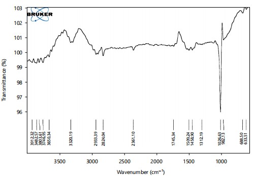
FTIR spectrum: The IR spectra were used to evaluate the existence of different chemical compounds in H. arcuatum based on peak values in the infra-red region. The methanol extract contained the groups like May 10, 2024C=C, C-H, C-O, O-H and N-H (Table 6; Fig. 5).
|
|
| Table 5: | GC-MS analysis of bioactive compounds of methanolic extracts of Hydrogonium arcuatum | |||
| RT | Area | Names of the compound | Molecular Formula |
| 9.93 | 13.23 | Caryophyllene | C15H24 |
| 16.84 | 62.48 | Diethyl phthalate | C12H14O4 |
| 20.57 | 12 | Benzenepropanoic acid,3,5-bis(1,1-dimethylethyl)-4- | C18H28O3 |
| 23.77 | 5.07 | hydroxyl-methyl ester Carotene,3,4-didehydro-1,2-dihydro-1-methoxy- |
C41H58O |
| 30.99 | 24.24 | Glycine, N-[(3a,5a)-24-oxo-3-[(trimethylsilyl)oxy]cholan-24-yl]-,methyl ester | C30H53NO4SI |
| 33 | 8.72 | 7,7,9,9,11,11-Hexamethyl-3,6,8,10,12,15-hexaoxa- | C14H36O6SI3 |
| 36.75 | 5.17 | 7,9,11-trisilaheptadecane 6,10,14-trimethyl-2-pentadecanone |
C18H36O |
|
| Table 6: | Detection of functional group in Hydrogonium arcuatum by FTIR | |||
| Frequency | Group | Appearance | Compound |
| 633.51 | C-Br stretching | Strong | Halo compound |
| 688.5 | C = C bending | Strong | Alkene |
| 983.13 | C = C bending | Strong | Alkene |
| 1026.63 | C-N stretching | Medium | Amine |
| 1312.19 | S = O stretching | Strong | Sulfone |
| 1458.9 | C-H bending | Medium | Alkene |
| 1509.24 | N-O stretching | Strong | Nitro-compound |
| 1745.34 | C = O stretching | Strong | Cyclopentanone |
| 2824.04 | C-H stretching | Medium | Aldehyde |
| 2933.39 | C-H stretching | Medium | Alkane |
| 3320.19 | N-H stretching | Medium | Secondary amine |
| 3650.34 | O-H stretching | Sharp | Alcohol |
Cytotoxicity assessment: Hydrogonium arcuatum extract exhibited the maximum cytotoxicity against HT- 29 but not against HCT-116 at a concentration of 1000 μg/mL and did not show any effect on both cell lines at doses of 50 and 100 μg/mL significantly (p<0.001) when compared to control (Fig. 6).
DISCUSSION
The phytochemical screening of a qualitative analysis of H. arcuatum revealed the presence of alkaloids, phenols and flavonoids in the methanolic extract and a moderate amount of carbohydrates, tannins, carbohydrates and terpenoids. The methanolic extract of H. arcuatum under investigation contained the highest level of total phenolic, flavonoid and alkaloid content. The over-expression of defense response genes to fight during stress makes it clear that phytoconstituents exhibit a shift in their quantities43. The results obtained by Karim et al.44 are in accordance with the current findings, where a high amount of TPC was observed in the methanol extract.
The antioxidant activity of the plant extract as IC50 value was determined to be high potential in methanol and low in n-hexane against DPPH and NOSA, respectively. As a result, numerous attempts to evaluate the pharmacological and nutraceutical potential of bryophytes have been made and it has been demonstrated that these amphibious plants are a priceless source of antioxidative, anti-cancerous, antibacterial and antiviral chemicals45.
The presence of caryophyllene, carotene, etc. was revealed by the GC-MS analysis. Caryophyllene, a phenol derivative is known to possess anti-inflammatory, antiviral, antifungal and anti-cancer properties. Several essential oils such as pepper and clove include caryophyllene, a naturally occurring bicyclic sesquiterpene in their formulation46. Caryophyllene is known to contain a rare cyclobutene ring which is an uncommon occurrence in the natural world.
The FTIR analysis of H. arcuatum methanol extract contained the groups like C=C, C-H, C=O, C-N, O-H and N-H. The peak at 1509 cm–1 showed the presence of nitro compounds and 1745 indicates the presence of cyclopentanone. Similar results were also reported by other researchers for the methanolic extract of moss47.
The H. arcuatum extract exhibited the maximum cytotoxicity against HT-29 but not against HCT-116 at a concentration of 1000 μg/mL and did not show any effect on both cell lines at doses of 50 and 100 μg/mL. Using the MTT method, the cytotoxicity potential of certain moss essential oils was evaluated in previous research on human immortalized keratinocytes, non-tumor cells and colorectal and breast tumor cells (HCT- 116 and MCF-7). Treatment with the sample essential oil in the different cell lines does not cause any harm in majority of the detected concentrations48.
Further investigation into the specific phytochemical constituents of H. arcuatum, particularly those responsible for its observed biological activities, could provide deeper insights into its potential therapeutic applications. Exploration of the mechanisms underlying the observed cytotoxicity against HT-29 cells could elucidate the pathways through which H. arcuatum exerts its anti-cancer effects. Comparative studies with other bryophytes or traditional medicinal plants may help identify unique bioactive compounds and broaden the understanding of H. arcuatum's pharmacological potential.
However, there are a few limitations of this study, such as, the study could benefit from additional experiments to validate the observed biological activities of H. arcuatum, such as in vivo studies or further cell line investigations. The cytotoxic effects observed against HT-29 cells need to be confirmed through additional assays and exploration of potential mechanisms of action. The extrapolation of the findings to clinical applications may be limited due to the use of cell-based assays and the absence of in vivo studies, warranting caution in interpreting the potential therapeutic relevance of H. arcuatum.
CONCLUSION
The present study was carried out on Hydrogonium arcuatum. Phytochemical screening revealed that the methanolic extract of H. arcuatum contains mainly alkaloids, phenols and flavonoids. The non- enzymatic assays DPPH and NOSA were used to evaluate the antioxidant capabilities of H. arcuatum. The primary objective of the DPPH assay is to assess the antioxidant potential of plant extracts including methanol and ethyl acetate. The GC-MS, FTIR and TLC were used to estimate the bioactive chemicals in a plant extract. Cytotoxicity assessment was done to investigate the effects on HT-29 and HCT-116 colorectal. It was found that H. arcuatum showed a highly significant increase in the cytotoxicity against HT-29 cell lines at 1000 concentration, significantly (p<0.001) when compared to control. The use of these bryophytes may be thought of as a useful method for treating colorectal cancer due to their cytotoxic activity, which lowers disease recurrence and can be used to create new drugs for a range of diseases in the pharmaceutical sector. The findings of this study suggest that bryophytes are abundant in phytoconstituents with significant bioactivities, including the potential for anti-proliferative effects. It is possible, however, that these will be useful in the future for other drug therapies for cancer cell lines.
SIGNIFICANCE STATEMENT
This study was aimed at unraveling the anti-oxidant potential, phytochemical profile and anti-cancerous activity of Hydrogonium arcuatum to highlight the rich phytochemical constituents of mosses. Scientists nowadays are looking for natural plant-based treatment options and phytochemicals from bryophytes can provide the much-needed alternative to chemically synthesized medicines. In the present study, extracts of H. arcuatum formulated in different solvents were subjected to GC-MS, FTIR and TLC procedures. In the phytochemical screening, methanolic extracts revealed the presence of tannins, alkaloids, flavonoids and phenols. The GC-MS analysis confirmed the presence of Caryophyllene, a potentially anti-viral, anti-inflammatory and cytotoxic compound. The significant cytotoxic activity of methanolic extract of H. arcuatum suggested its potential in the treatment of colorectal cancer.
REFERENCES
- Singh, D.K. and D. Singh, 2016. Epiphyllous liverworts of India: An overview. Plant Sci. Today, 3: 157-174.
- Kutnar, L., J. Kermavnar and M.S. Sabovljević, 2023. Bryophyte diversity, composition and functional traits in relation to bedrock and tree species composition in close-to-nature managed forests. Eur. J. For. Res., 142: 865-882.
- Shaw, J. and K. Renzaglia, 2004. Phylogeny and diversification of bryophytes. Am. J. Bot., 91: 1557-1581.
- Beckert, S., H. Muhle, D. Pruchner and V. Knoop, 2001. The mitochondrial nad2 gene as a novel marker locus for phylogenetic analysis of early land plants: A comparative analysis in mosses. Mol. Phylogenet. E, 18: 117-126.
- Flores-Sandoval, E., R. Nishihama and J.L. Bowman, 2024. Hormonal and genetic control of pluripotency in bryophyte model systems. Curr. Opin. Plant Biol., 77.
- Srivastava, S.C. and K.K. Rawat, 2001. On a long-lost endemic liverwort (Hepaticae) from India. Curr. Sci., 80: 1484-1486.
- Mishra, M., P.K. Dash, A. Alam, S. Sahoo and R. Das, 2016. Current status of diversity and distribution of Bryophytes of Odisha. Plant Sci. Today, 3: 186-194.
- Singh, M., A.K. Rawat and R. Govindarajan, 2007. Antimicrobial activity of some Indian mosses. Fitoterapia, 78: 156-158.
- Alam, A., K.K. Rawat, P.K. Verma, V. Sharma and D.S. Gupta, 2015. Moss flora of central India. Plant Sci. Today, 2: 159-171.
- Rawat, K.K., V. Sahu and R.R. Paul, 2021. Bryophytes of Mount Abu, Rajasthan, India. Nelumbo, 63: 207-217.
- Flowers, S., 1957. Ethnobryology of the gosuite Indians of Utah. Bryologist, 60: 11-14.
- Saxena, D.K. and Harinder, 2004. Uses of bryophytes. Resonance, 1: 56-65.
- Shaw, A.J., 2008. Bryophyte Species and Speciation. In: Bryophyte Biology, Shaw, A.J. (Ed.), Cambridge University Press, Cambridge, England, ISBN: 9780511754807, pp: 445-486.
- Asakawa, Y., M. Toyota, T. Takemoto and R. Mues, 1981. Aromatic esters and terpenoids of the liverworts in the genera Trichocolea, Neotrichocolea and Trichocoleopsis. Phytochemistry, 20: 2695-2699.
- Alam, A., 2012. Some Indian bryophytes known for their biologically active compounds. Int. J. Appl. Biol. Pharm. Technol., 3: 239-246.
- Asakawa, Y., 1995. Chemical Constituents of the Bryophytes. In: Progress in the Chemistry of Organic Natural Products, Asakawa, Y. (Ed.), Springer, Vienna, Austria's, ISBN: 978-3-7091-6896-7, pp: 1-562.
- Asakawa, Y., F. Nagashima, T. Hashimoto, M. Toyota and A. Ludwiczuk et al., 2014. Pungent and bitter, cytotoxic and antiviral terpenoids from some bryophytes and inedible fungi. Nat. Prod. Commun., 9.
- Asakawa, Y., M. Toyota, M. Tori and T. Hashimoto, 2000. Chemical structures of macrocyclic bis(bibenzyls) isolated from liverworts (Hepaticae). J. Spectrosc., 14: 149-175.
- Lu, R., C. Paul, S. Basar and W.A. König, 2005. Sesquiterpene constituents of the liverwort Lophozia ventricosa. Tetrahedron: Asymmetry, 16: 883-887.
- Chopra, R.N. and P.K. Kumra, 1988. Biology of Bryophytes. Wiley, New York, ISBN: 9780852262405, Pages: 350.
- Jocković , N., P.B. Andrade, P. Valentão and M. Sabovljević, 2008. HPLC-DAD of phenolics in bryophytes Lunularia cruciata, Brachytheciastrum velutinum and Kindbergia praelonga. J. Serb. Chem. Soc., 73: 1161-1167.
- Sabovljevic, A., M. Sokovic, J. Glamolija, A. iri, M. Vujii, B. Pejin and M. Sabovljevi, 2011. Bio-activities of extracts from some axenically farmed and naturally grown bryophytes. J. Med. Plants Res., 5: 565-571.
- Adebiyi, A.O., A.A. Oyedeji, E.E. Chikwendu and O.A. Fatoke, 2012. Phytochemical screening of two tropical moss plants: Thidium gratum P. Beauv and Barbula indica brid grown in Southwestern Ecological Zone of Nigeria. Am. J. Anal. Chem., 3: 836-839.
- Nordberg, J. and E.S.J. Arnér, 2001. Reactive oxygen species, antioxidants, and the mammalian thioredoxin system. Free Radic. Biol. Med., 31: 1287-1312.
- Kris-Etherton, P.M., K.D. Hecker, A. Bonanome, S.M. Coval and A.E. Binkoski et al., 2002. Bioactive compounds in foods: Their role in the prevention of cardiovascular disease and cancer. Am. J. Med., 113: 71-88.
- Cai, H. and D.G. Harrison, 2000. Endothelial dysfunction in cardiovascular diseases: The role of oxidant stress. Circ. Res., 87: 840-844.
- Behera, B.C., N. Verma, A. Sonone and U. Makhija, 2006. Determination of antioxidative potential of lichen Usnea ghattensis in vitro. LWT-Food Sci. Technol., 39: 80-85.
- Vats, S. and A. Alam, 2013. Antibacterial activity of Atrichum undulatum (Hedw.) P. Beauv. against some pathogenic bacteria. J. Biol. Sci., 13: 427-431.
- Bhattarai, H. D., B. Paudel, H.K. Lee, H. Oh and J.H. Yim, 2009. In vitro antioxidant capacities of two benzonaphthoxanthenones: Ohioensins F and G, isolated from the Antarctic moss Polytrichastrum alpinum. Z. Naturforsch C, 64: 197-200.
- Asakawa, Y., 2007. Biologically active compounds from bryophytes. Pure Appl. Chem., 79: 557-580.
- Shi, L., W. Zhao, Z. Yang, V. Subbiah and H.A.R. Suleria, 2022. Extraction and characterization of phenolic compounds and their potential antioxidant activities. Environ. Sci. Pollut. Res., 29: 81112-81129.
- Asakawa, Y. and A. Ludwiczuk, 2013. Bryophytes: Liverworts, Mosses, and Hornworts: Extraction and Isolation Procedures. In: Metabolomics Tools for Natural Product Discovery: Methods and Protocols, Roessner, U. and D.A. Dias (Eds.), Humana Press, Totowa, New Jersey, ISBN: 978-1-62703-577-4, pp: 1-20.
- Harborne, J.B., 1998. Phytochemical Methods a Guide to Modern Techniques of Plant Analysis. Springer, Netherlands, Pages: 302.
- Bhadauriya, G., K.S. Rathore and S. Singh, 2018. Phytochemical screening and total phenolic content in the extract of bryophyte Plagiochasma appendiculatum and Dicranum scoparium. Environ. Conserv. J., 19: 175-181.
- Khanal, L.N., K.R. Sharma, Y.R. Pokharel and S.K. Kalauni, 2022. Phytochemical analysis and in vitro antioxidant and antibacterial activity of different solvent extracts of Beilschmiedia roxburghiana Nees stem barks. Sci. World J., 2022.
- Madaan, R., G. Bansal, S. Kumar and A. Sharma, 2011. Estimation of total phenols and flavonoids in extracts of Actaea spicata roots and antioxidant activity studies. Indian J. Pharm. Sci., 73: 666-669.
- Abu Bakar, M.F., F. Abdul Karim, M. Suleiman, A. Isha and A. Rahmat, 2015. Phytochemical constituents, antioxidant and antiproliferative properties of a liverwort, Lepidozia borneensis stephani from Mount Kinabalu, Sabah, Malaysia. Evidence-Based Complementary Altern. Med., 2015.
- Asakawa, Y., K. Nii and M. Higuchi, 2015. Identification of sesquiterpene lactones in the Bryophyta (mosses) Takakia: Takakia species are closely related chemically to the Marchantiophyta (liverworts). Nat. Prod. Commun., 10.
- Pejin, B., J. Bogdanovic-Pristov, I. Pejin and M. Sabovljevic, 2013. Potential antioxidant activity of the moss Bryum moravicum. Nat. Prod. Res., 27: 900-902.
- Badami, S., S. Moorkoth, S.R. Rai, E. Kannan and S. Bhojraj, 2003. Antioxidant activity of Caesalpinia sappan heartwood. Biol. Pharm. Bull., 26: 1534-1537.
- Shamran, D.J. and E.F. Al-Jumaili, 2020. Phytochemical screening by HPLC and FTIR spectroscopy of Glucokinin isolated from methanol extract of Bauhinia variegata. Medico Legal Update, 20: 759-763.
- Ganji, P.N., W. Park, J. Wen, H. Mahaseth and J. Landry et al., 2013. Antiangiogenic effects of ganetespib in colorectal cancer mediated through inhibition of HIF-1α and STAT-3. Angiogenesis, 16: 903-917.
- Isah, T., 2019. Stress and defense responses in plant secondary metabolites production. Biol. Res., 52.
- Karim, F.A., M. Suleiman, A. Rahmat and M.F. Abu Bakar, 2014. Phytochemicals, antioxidant and antiproliferative properties of five moss species from Sabah, Malaysia. Int. J. Pharm. Pharm. Sci., 6: 292-297.
- Alam, A., 2021. Potential of bryophytes in prevention and medication of COVID-19. Ann. Phytomed., 10: S121-S129.
- Sheffield, L. and J. Rowntree, 2009. Bryophyte biology, 2nd edn. Ann. Bot., 104.
- Mohandas, G.G. and M. Kumaraswamy, 2018. Antioxidant activities of terpenoids from Thuidium tamariscellum (C. Muell.) Bosch. and Sande-lac. a moss. Pharmacogn. J., 10: 645-649.
- Klegin, C., N.F. de Moura, M.H.O. de Sousa, R. Frassini and M. Roesch‐Ely et al., 2021. Chemical composition and cytotoxic evaluation of the essential oil of Phyllogonium viride (Phyllogoniaceae, Bryophyta). Chem. Biodivers., 18.
How to Cite this paper?
APA-7 Style
Sharma,
T., Singh,
S., Alam,
A. (2024). Phytochemical Profiling, Antioxidant and Anti-Cancerous Activity of Hydrogonium arcuatum (Griff.) Wijk. & Marg. (Bryophyta: Pottiaceae). Asian Journal of Biological Sciences, 17(3), 469-481. https://doi.org/10.3923/ajbs.2024.469.481
ACS Style
Sharma,
T.; Singh,
S.; Alam,
A. Phytochemical Profiling, Antioxidant and Anti-Cancerous Activity of Hydrogonium arcuatum (Griff.) Wijk. & Marg. (Bryophyta: Pottiaceae). Asian J. Biol. Sci 2024, 17, 469-481. https://doi.org/10.3923/ajbs.2024.469.481
AMA Style
Sharma
T, Singh
S, Alam
A. Phytochemical Profiling, Antioxidant and Anti-Cancerous Activity of Hydrogonium arcuatum (Griff.) Wijk. & Marg. (Bryophyta: Pottiaceae). Asian Journal of Biological Sciences. 2024; 17(3): 469-481. https://doi.org/10.3923/ajbs.2024.469.481
Chicago/Turabian Style
Sharma, Tripti, Swati Singh, and Afroz Alam.
2024. "Phytochemical Profiling, Antioxidant and Anti-Cancerous Activity of Hydrogonium arcuatum (Griff.) Wijk. & Marg. (Bryophyta: Pottiaceae)" Asian Journal of Biological Sciences 17, no. 3: 469-481. https://doi.org/10.3923/ajbs.2024.469.481

This work is licensed under a Creative Commons Attribution 4.0 International License.